Report 85: “The Underlying Pathology of Spike Protein Biodistribution in People That Died Post COVID-19 Vaccination” – Dr. Arne Burkhardt
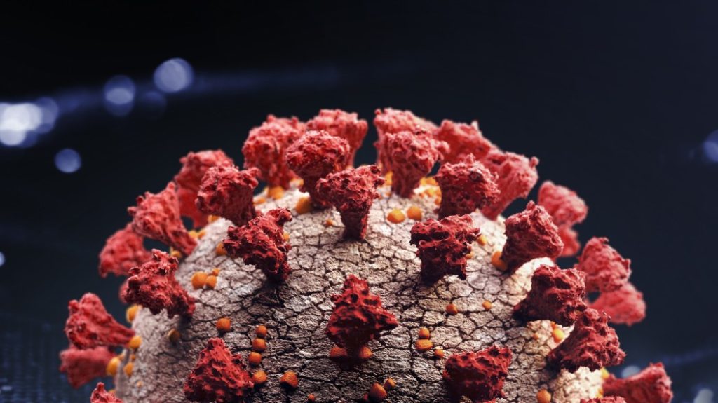
The Scientific and Medical Advisory Committee hosted a meeting with special guest speaker Professor Arne Burkhardt.
Dr. Arne Burkhardt
Compiled and edited by
Robert W. Chandler MD MBA*
*A transcript was produced then edited with a diligent effort to leave the meaning unchanged. My additions are in italics.
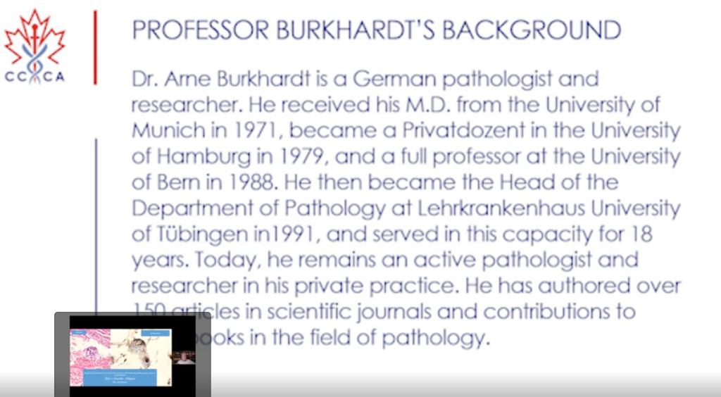
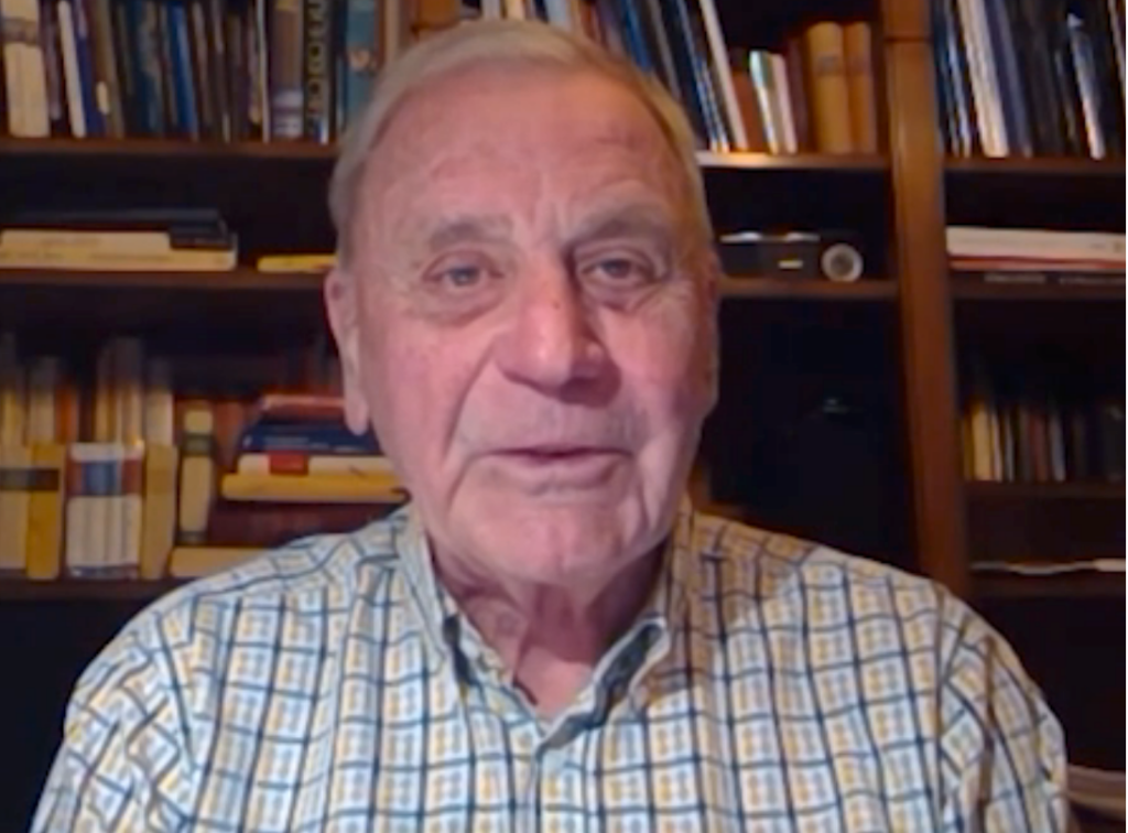
Professor Burkhardt was a German pathologist and researcher. He presented the findings of the ongoing work of an international team of pathologists. They have reviewed pathology specimens from people with new onset of symptoms or who have died following COVID-19 genetic vaccinations. He explained how to differentiate damage following natural infection with that following vaccination. Additionally, he presented tissue samples to illustrate the distribution of the spike protein following COVID-19 genetic vaccination and its associated damage, amyloid production, and clot formations.
Original Lecture
Transcript
I will tell you that actually very soon after the vaccine campaign started in Germany, there were relatives of deceased persons who went to me and said, well, we want to, we want that a pathologist takes a second opinion. We don’t believe that our relative died of natural causes. And I thought this was an easy task.
I said, well, of course, I will look at the slides or the specimens that were taken during autopsy, and I will give a second opinion. But very soon with the first three or four cases, I realized that this was really a task that I could not handle just by myself alone. And fortunately, I found a pathologist who worked with me, Professor Walter Lang.
He joined me in these endeavors and very soon we got, many more scientists and some pathologists, especially from outside of Germany, who joined us in these endeavors. And if I can show my first slide now. So, at this moment we are actually an international team.
It’s about 10 pathologists, coroners, biologists, chemists, and physicists, not only from Germany, but actually many European countries, especially Austria, Switzerland, Italy, and Sweden.
We have completed the review of 75 autopsy studies, and we are right now having 41 biopsies from living persons. There are 40 men, 35 women of the deceased. The age range was from 21 to 94 ages.
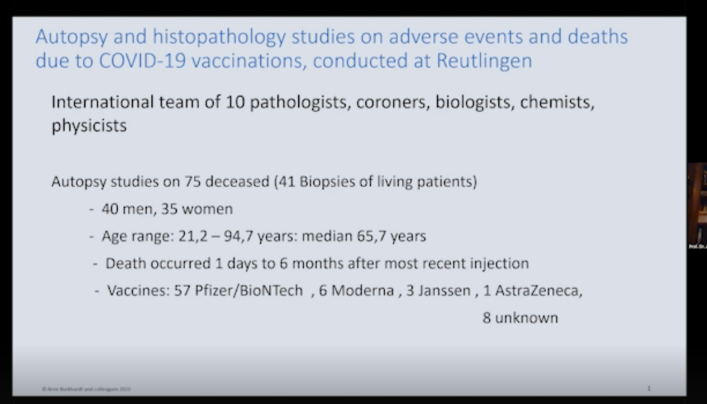
Deaths occurred one day or to six months after the most recent injection. And the vaccines are those that are commonly used in Germany. Pfizer-BioNTech vaccine was the most common, but sometimes it was in connection with other vaccinations.
The autopsies were done by pathologists, by coroners, and one by both. So, you see it’s about equal distribution. The primary diagnosis was natural death in 63 cases. And, in five cases, it was termed uncertain. So that makes 91% not related to the vaccination.
Only in seven cases it was stated that there might be a correlation with the vaccination and the disease. We did the second opinion, and these are our results. I will show you histological findings.
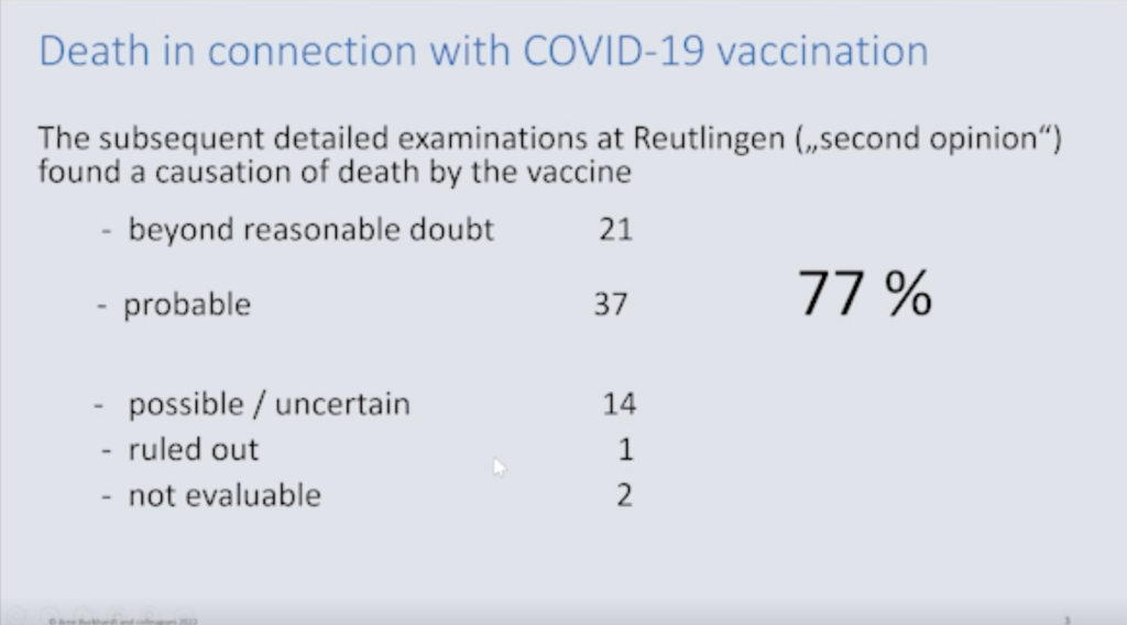
We saw a correlation of vaccination and the death occurrence in 21 cases, and in 37 cases it was probable. So, that is 77%. In another 14 (19%), we said it was uncertain or possible, ruled out in only one case, and two cases were not evaluable.
Sudden Adult Death Syndrome
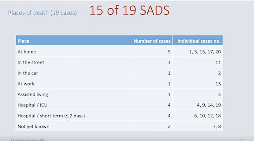
This is the evaluation of 19 cases where they died. This is one important thing that most of the diseased we evaluated died suddenly, mostly at home, in the street, in the car, at work. So, we can rule out any post-mortem changes of the organs. There’s no artificial respiration or anything like that.
So, of these 19 cases, 15 (79%) fulfill the criteria for what is now called the Sudden Adult Death Syndrome. And this is very important because I will come to the possible causes of this syndrome later.
COVID-19 vs. Modified mRNA (modRNA)
Now, at the beginning it was clear that there is a difference between the natural COVID-19 infection and the messenger modified RNA vaccination, but in both sets a spike protein that is probably the most important action that does harm to the tissues.
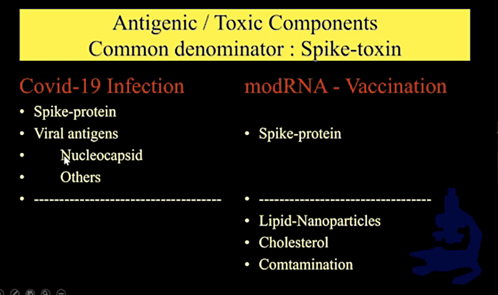
Now the COVID-19 infection, of course, has, besides a spike protein, other antigenic and possibly toxic agents like the nucleocapsid and others, and in the vaccination beside the spike protein, which of course is the leading mechanism. We have the lipid nanoparticles, we have cholesterol, and in some cases, contamination.
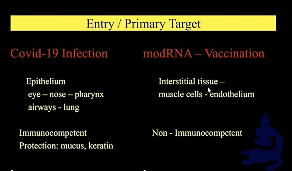
From the beginning, we realized that there’s a different entry or primary target of these toxic and antigenic agents and the infection. It’s the epithelium, starting from the nose, from the eye, pharynx, airways, lung. And these are immunocompetent linings of the body. And also, there’s some protection by mucus and keratin.
Now, in the vaccination we do not use this natural entrance, but it is directly shot into the interstitial tissue, the basal tissue and the endothelium. And these are non-immunocompetent.
Methodology
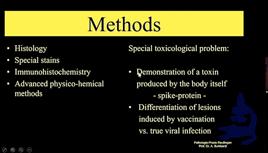
We used normal histology (hematoxylin and eosin stain), special stains, immunohistochemistry, and in some cases advanced physical chemical methods.
From the beginning on it was clear to us that this was a novel examination because we had to demonstrate a toxin that was produced by the body itself, the spike protein. And this had to be differentiated from lesions induced by true viral infections.
And, as the body produces these toxins, we have to look for the toxin in the organs and tissues of the organ itself, which produce it, and not in body fluids or in the gastric contents.
Immunohistochemistry: Spike (COVID-19 + modRNA) vs. Nucleocapsid (COVID-19)
So, we applied the method of immunochemistry to show the spike protein on the one hand and nucleocapsid on the other hand. Maybe most of you probably know the mechanism of immunochemistry is to have some stained material that binds to the protein that you are looking for. You see here some positively stained cells for spike protein from a nasal swab.
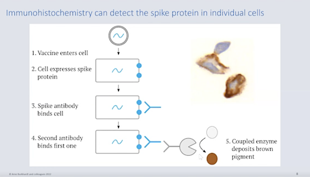
And this method was used to demonstrate the spike protein and, on the other hand, in some cases we excluded or found nucleocapsid.
General Lesions Affecting More Than One Organ (See Supplemental Resources SR 1- SR 5.)
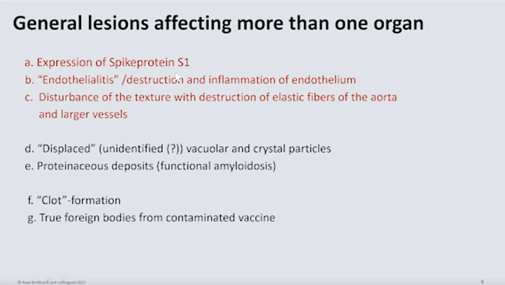
First of all, we have general lesions affecting more than one organ. And, this is the expression of the spike protein S1. (See SR 1 and SR 2.)
Then, we have especially the detrimental causes on the vessels because, no matter how you inject the vaccine, it will always go into the blood and lymph vessels. We found what is called an endotheliitis. It’s an inflammation and destruction of endothelium. And also, the larger vessels and the aorta are an aim of this toxic action. It’s a disturbance of the texture and the destruction of elastic fibers in the larger vessels.
When we started, it was put forward by the pharmaceutical companies that it stays in the deltoid muscles where it was injected and that the muscle cells and maybe some other interstitial cells would produce the spike protein and cause the immunological reaction that they planned to.
Expression of Spike Protein
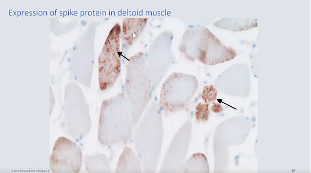
Here we examined the deltoid muscle in one case, and you can see this granular markation (black arrows) of the muscle cells. Spike protein was produced at the spot of injection.
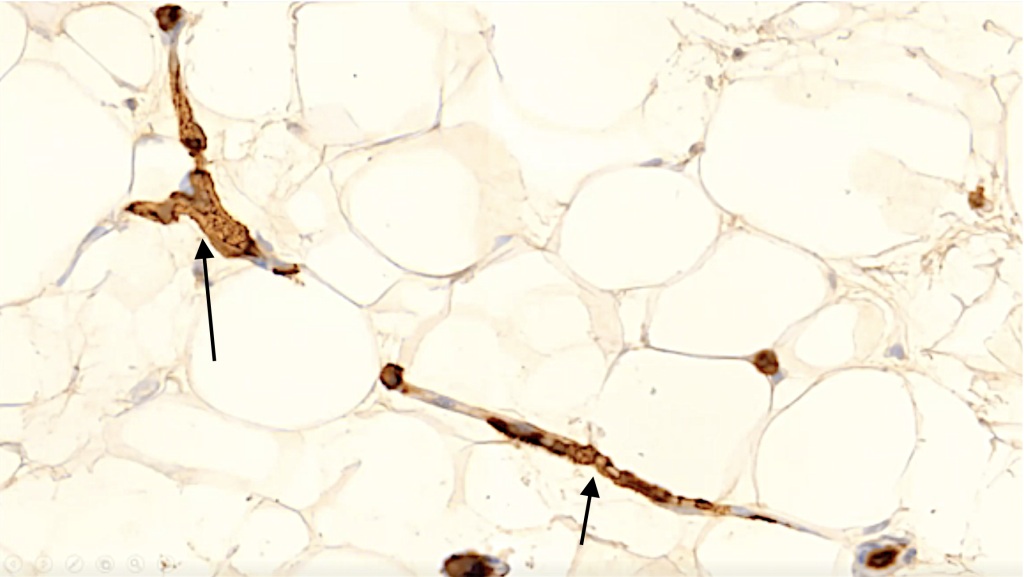
Then we also found it in capillaries (black arrows). Here you see in fat tissue, you see the vacuoles and you see this capillary. Here it is out of the planar section, and here you can see it is cut vertically and you can see the endothelium strongly expresses a spike protein.
Endotheliitis
The spike protein, as you know, as we know now, it’s a toxic agent and it harms the endothelium as we will see later.
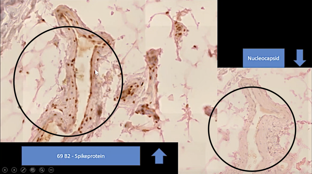
It is also found in other vessels in the surrounding, and you can see here an arteriole with a clear markation of the endothelium and some also in cells of the vessel walls. And this is a nucleocapsid stain of the same vessel. As you may see, it’s a completely negative reaction.
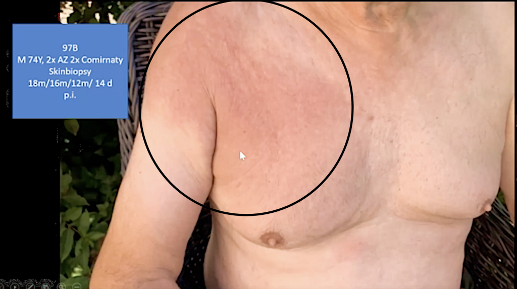
Here you can see at the site where the injection was performed. We can see still, for a very long time, inflammatory reaction in some cases. (See SR 3 and SR 4.)
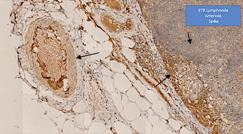
This is a biopsy from this person that you’ve just seen. You can see that in the lymph node a strong demonstration of the spike protein.
After that, the spike protein is distributed or can be demonstrated in many organs, almost all organs, more or less.
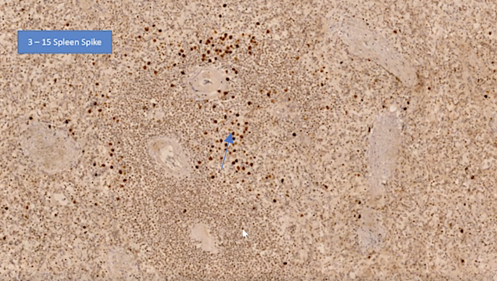
Spike in the spleen.
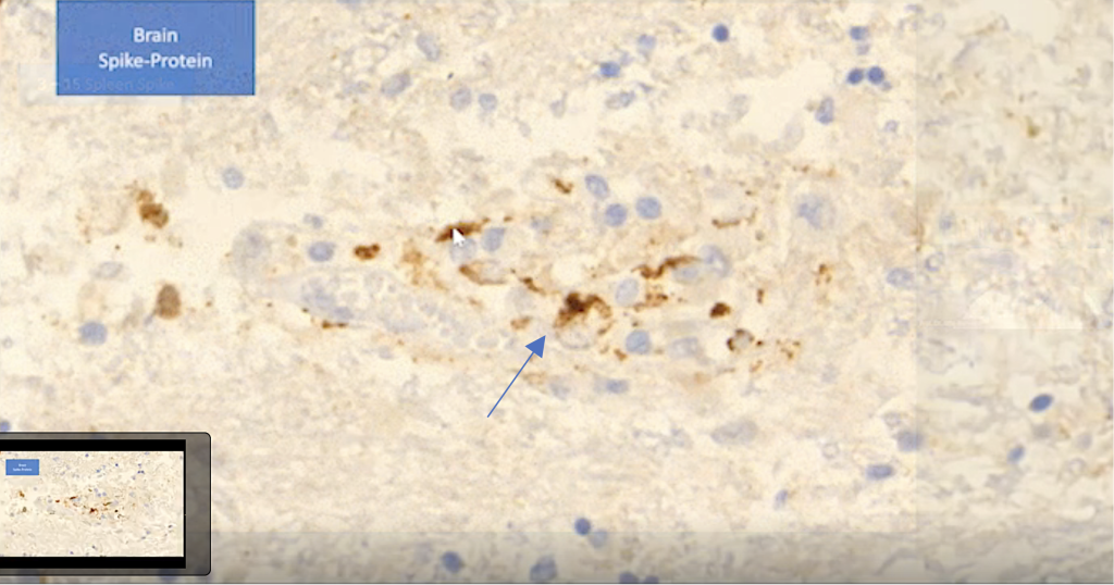
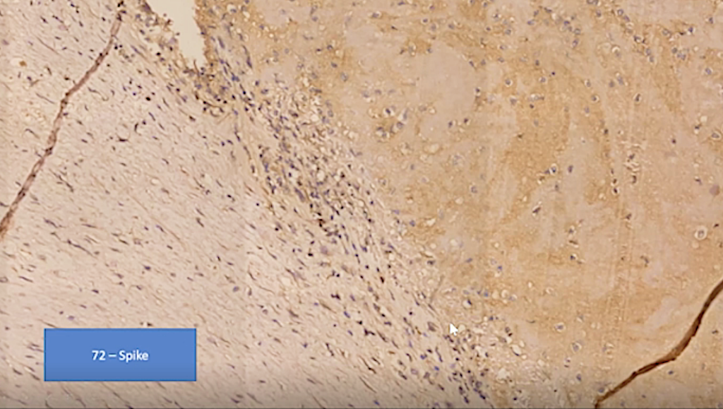
Spike protein in brain tissue.
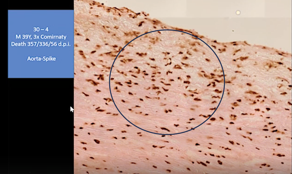
Spike protein in aorta.
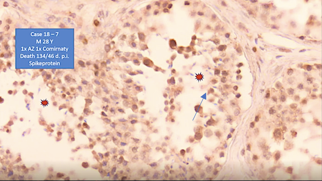
Spike in testis.
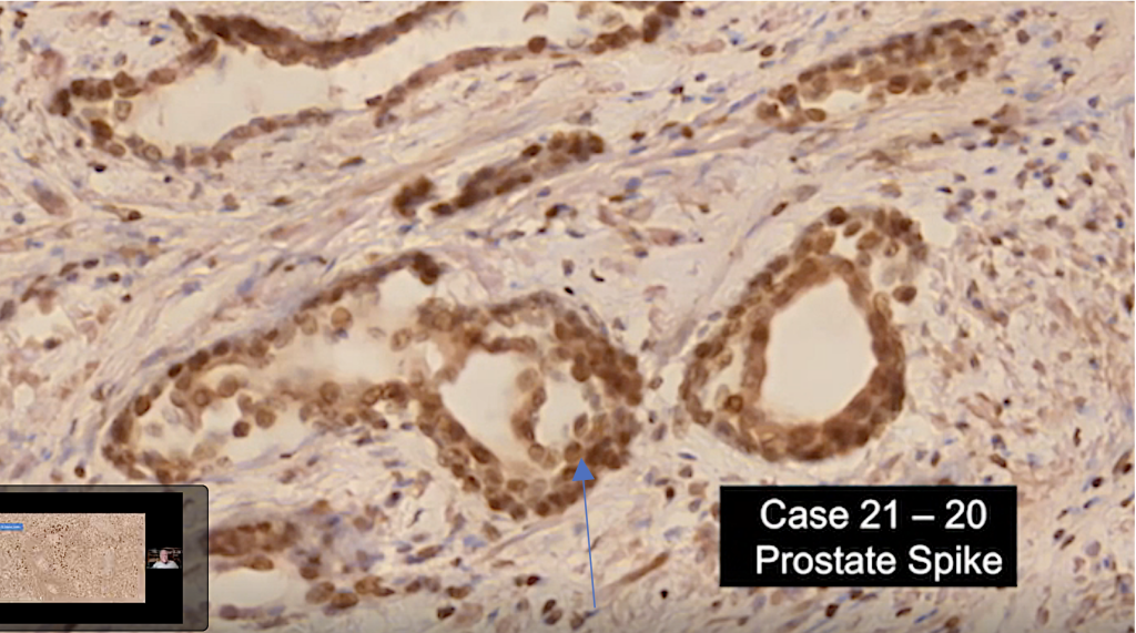
Spike in prostate.
There’s some publication now about the effects on male fertility.

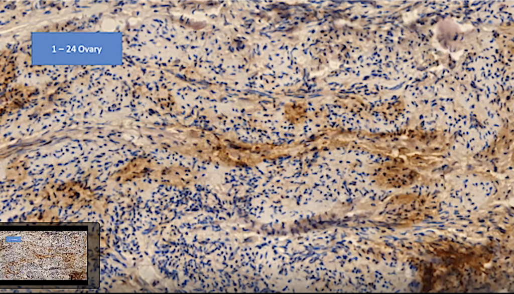
Post-modRNA lymphocytic infiltration in ovary.
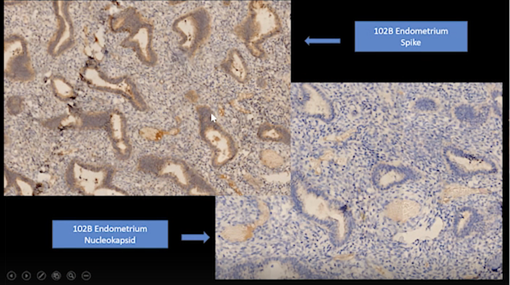
Post-modRNA lymphocytic infiltration in endometrium.
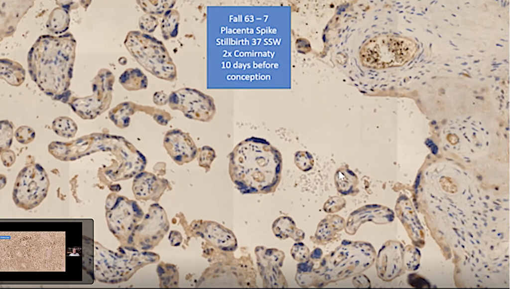
Post-modRNA lymphocytic infiltration in placenta.
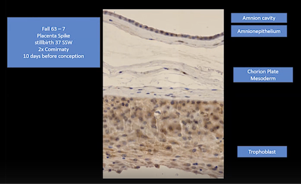
Post-modRNA lymphocytic infiltration in placenta and spike.
So, this is the first round about the general regions that we found.
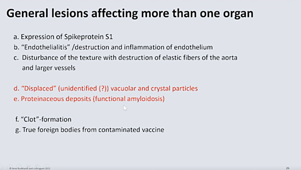
Displaced Unidentified Vacuolar and Crystal Particles
And we come now to the displaced vacuole and crystal particles at the proteinaceous deposits.
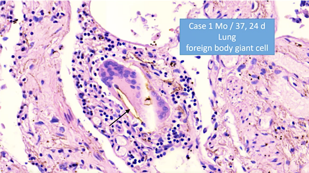
First of all, we found in some cases in the lung and also in other organs, these foreign body giant cells with these needle-like in inclusions (black arrow). Now we know these inclusions to be so-called ‘cholesterol needles.’
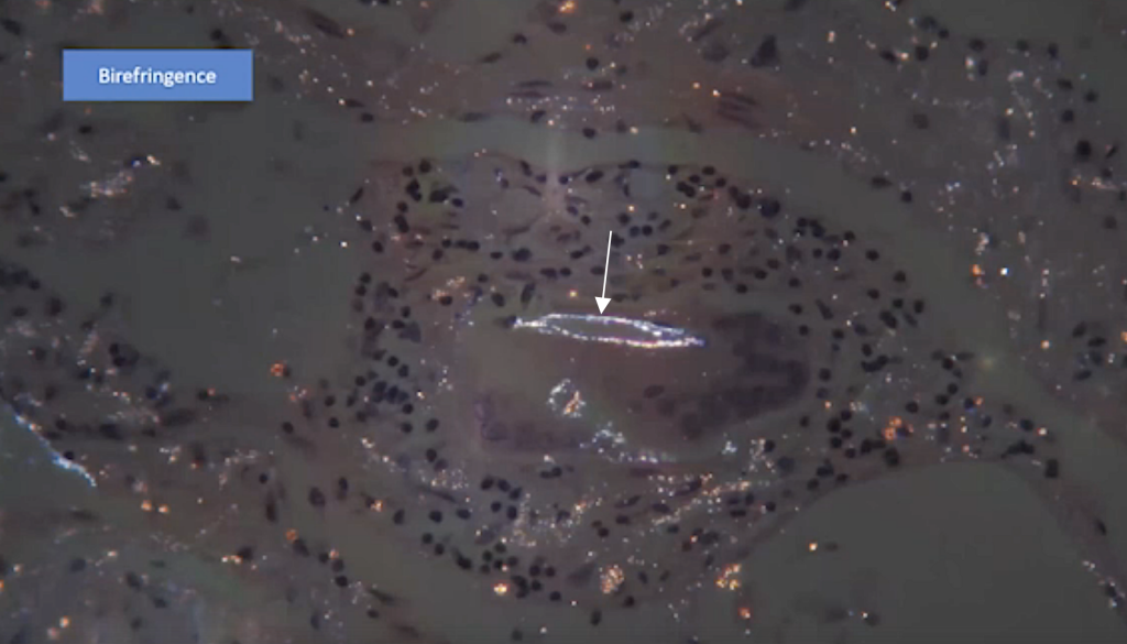
In addition, we found some other material there, and you can see that in the periphery you can see a birefringence of these materials (white arrow).
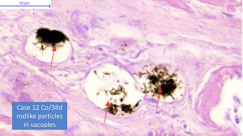
And then we found, in many organs, these rod-like particles (red arrows) which are in vacuoles. Now, these probably don’t form vacuoles, but they contained lipids, which like in the fat tissue are extracted during preparation.
First of all, we overlooked these particles, because we thought it was so called formalin pigment, which is very well known to pathologists to be an artifact.
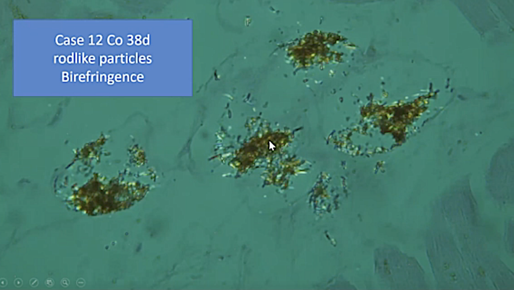
But we came to the conclusion that this is definitely not an artifact but that they must be some kind of material which is formed in the process of this vaccination. You can see some of these are also birefringent (white arrow).
We thought a lot about where this comes from. It could come from the injection itself, but the mass that we saw was not compatible.
It’s just I think it would not be possible that so much material is injected.
Also, the production of some substances could be the cause for these; but from, Raman Spectrometry, we had the notion that it could be cholesterol. And then we figured out that where cholesterol is located in the human body.
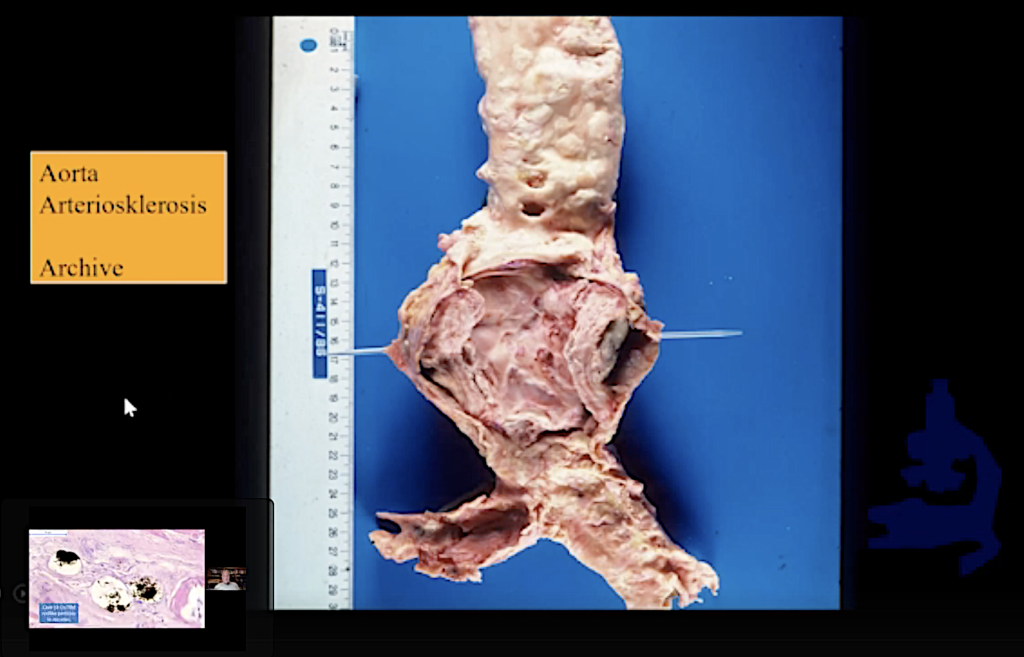
And, as you may know, now, this is not a vaccination victim. This is a normal person who died of natural causes, but he had this large arteriosclerotic plaque with perforation, and this contains, of course, masses of cholesterol.
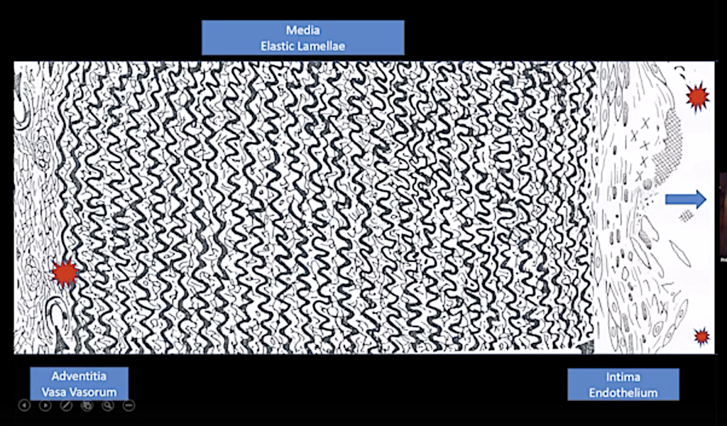
And just to show you the scheme of the aorta — you can see that the atheroma contain these needle-like cholesterols. And, if the epithelium is destroyed, it might come into the blood circulation. (Cholesterol embolus after erosion of the inner lining of the artery overlying the plaque.)
And secondary destruction could be due to the endothelial lesions of the vasa vasorum.
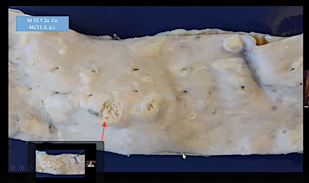
And here you can see a 55-year-old man, the aorta, and you can see that he has some atheromatous plaques; and you can see that one of them has broken open (red arrow), and the contents of these atheromatous plugs went into the blood.
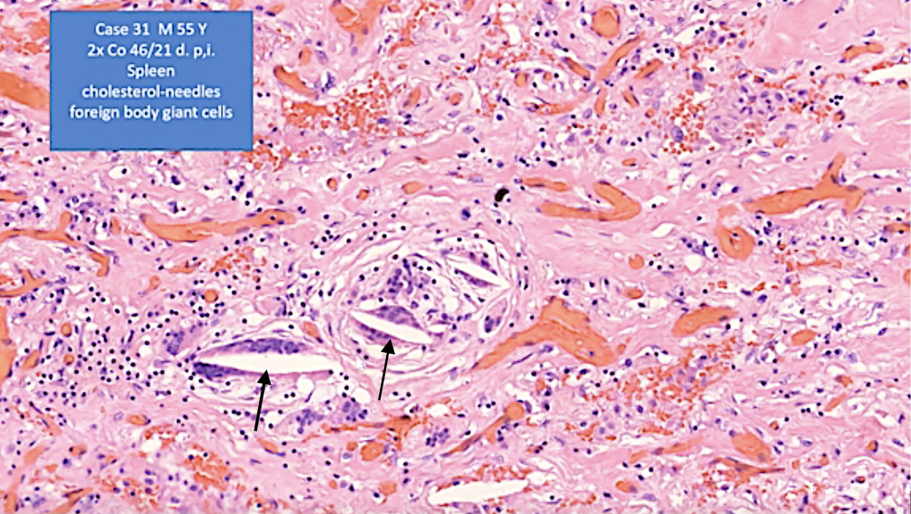
And we can show this because, in the spleen of this person, we found these foreign body giant cells with cholesterol needles (black arrows), and this seems to be very reasonable that this is one of the sources of cholesterol in the in the organs of the deceased after vaccination.
Amyloid Formation (See SR 6.)
We come to the so-called ‘amyloid formation.’
It was Swedish authors which found that the peptide sequences of the spike protein might be similar to amyloid and that amyloid-like substances may be formed.

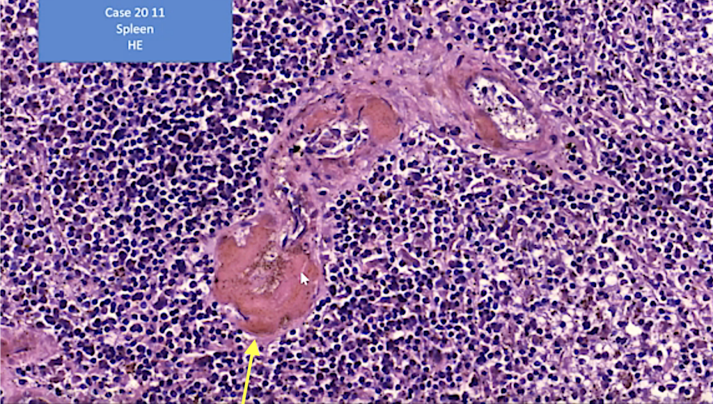
(The yellow arrow points to amyloid in the vessel wall.)
Very early we recognized that here was a strange extracellular deposit of strongly eosinophilic proteinaceous material in the vessel walls of many of the deceased that we looked at.
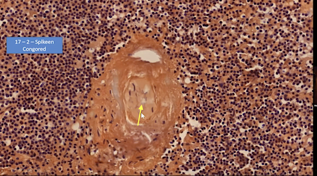
And, in some cases, even the vessel wall, here in the spleen, was occluded, and with the Congo Red stain these vessel walls are strongly positive.
So, we confirmed that this is an amyloid deposition (yellow arrow).
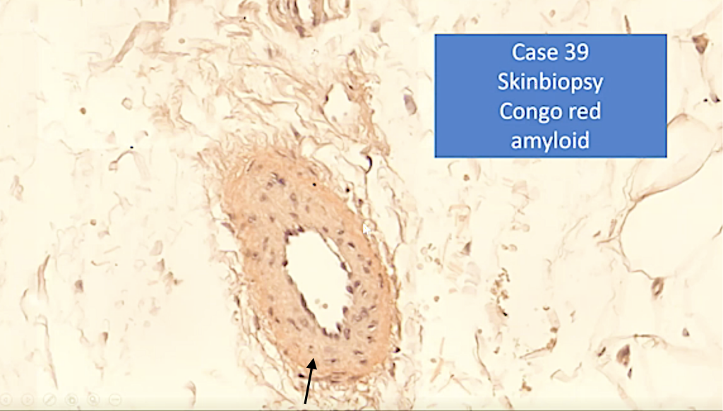
And this is a skin biopsy from a living person. She had problems of perfusion, blood perfusion. You can see also that a small subcutaneous vessel has some amyloid-like material (black arrow).
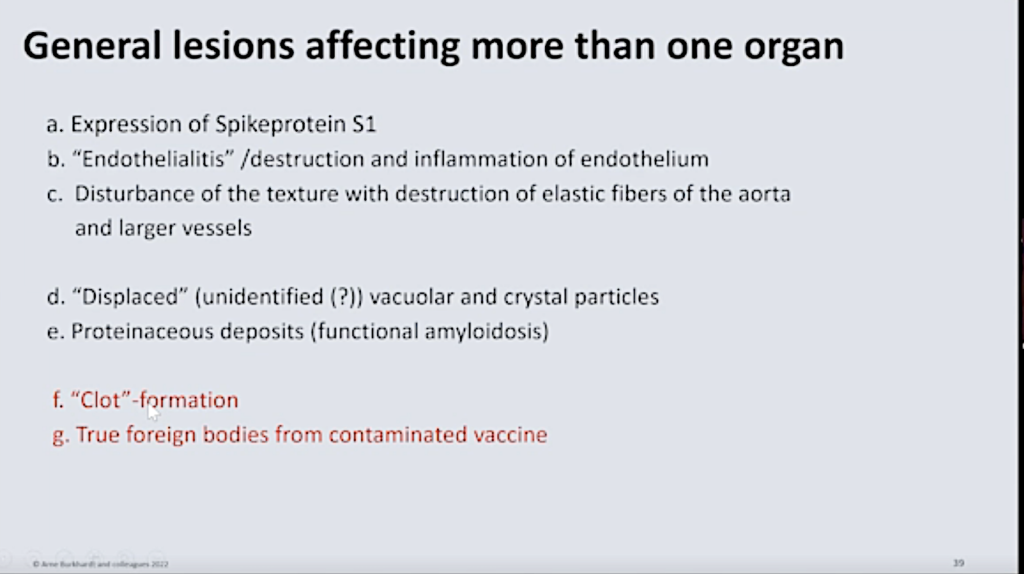
Clot Formation
Now we come to the so-called ‘clot formation.’
You may all be aware of these reports from undertakers that they found something that they never saw before; that there were some clots that were of elastic property.
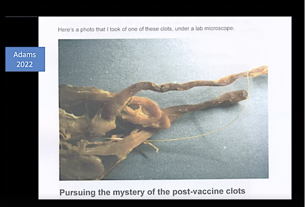
And, the only thing they said is that it was not a thrombus, but some (inaudible). That’s why they are called it a clot.
And, until now, we do not know exactly what it is, but it’s definitely not a thrombotic event. Because no person would survive these clot formations.
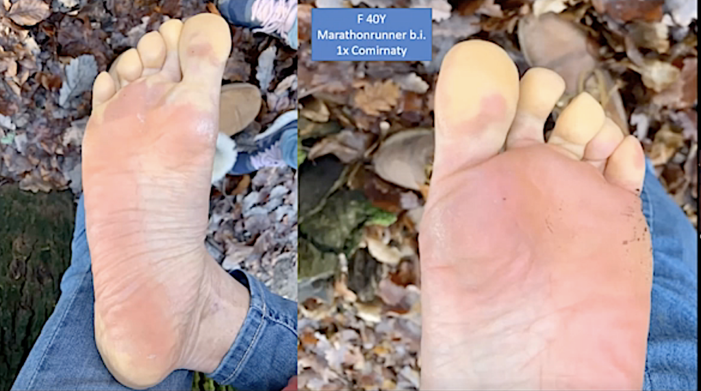
But we have a very interesting observation. We have this 40-year-old woman of around 40, which was an active marathon runner.
And after one vaccination with Comirnaty, she was hardly able to walk because she had this very severe disturbance of a perfusion of her lower legs.
And, in the radiograph, there was a dissection of the arteries of the lower leg.
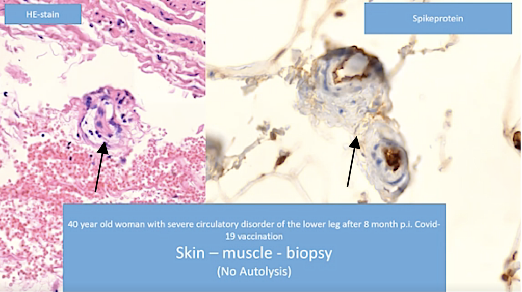
And, in the skin biopsy that was taken from her, we found these lesions of the small vessels, and you can see that the endothelium is swollen. It is detached from the basal membrane. And in these vessels, we could clearly demonstrate the spike protein.
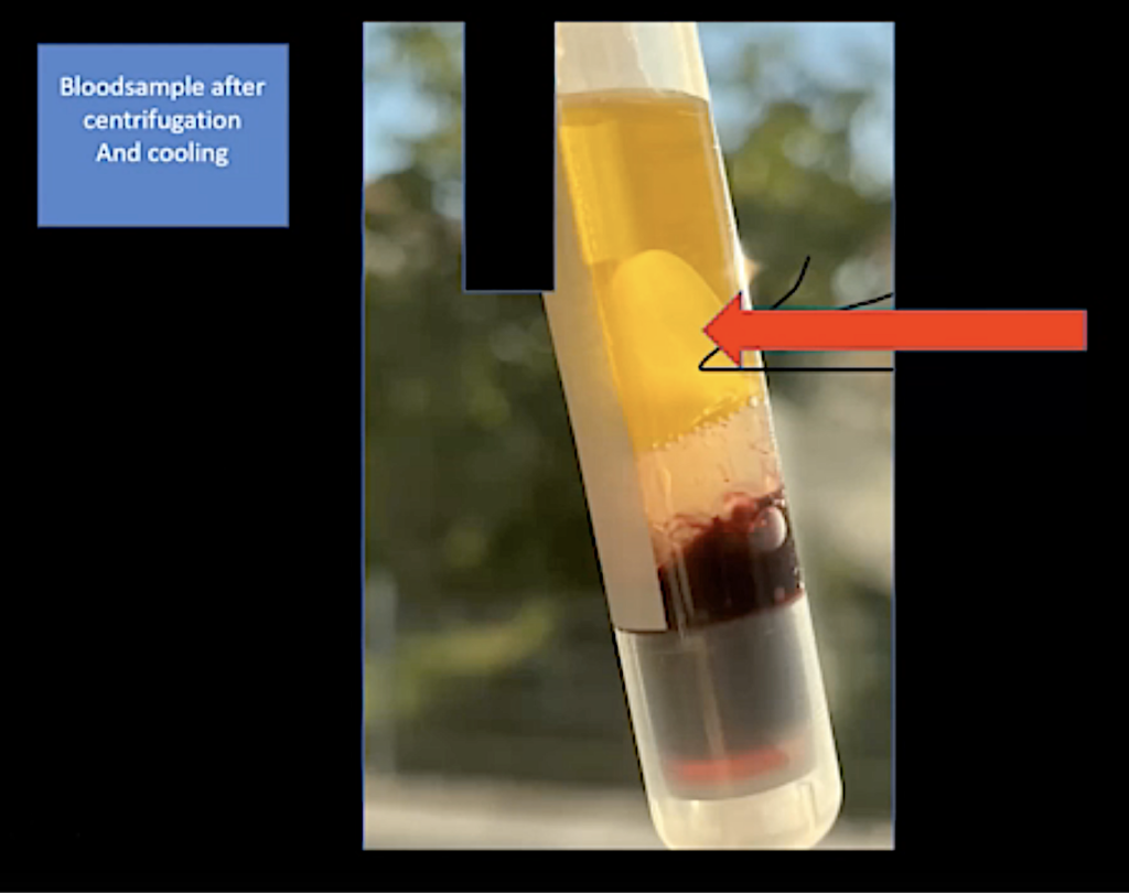
And this is from a living person. It’s definitely not an autolytic artifact. And, in this person, and this is the interesting part of it, blood samples were taken; and, after centrifugation and cooling, these clots were formed. There they are definitely separated from this part of the centrifugation.
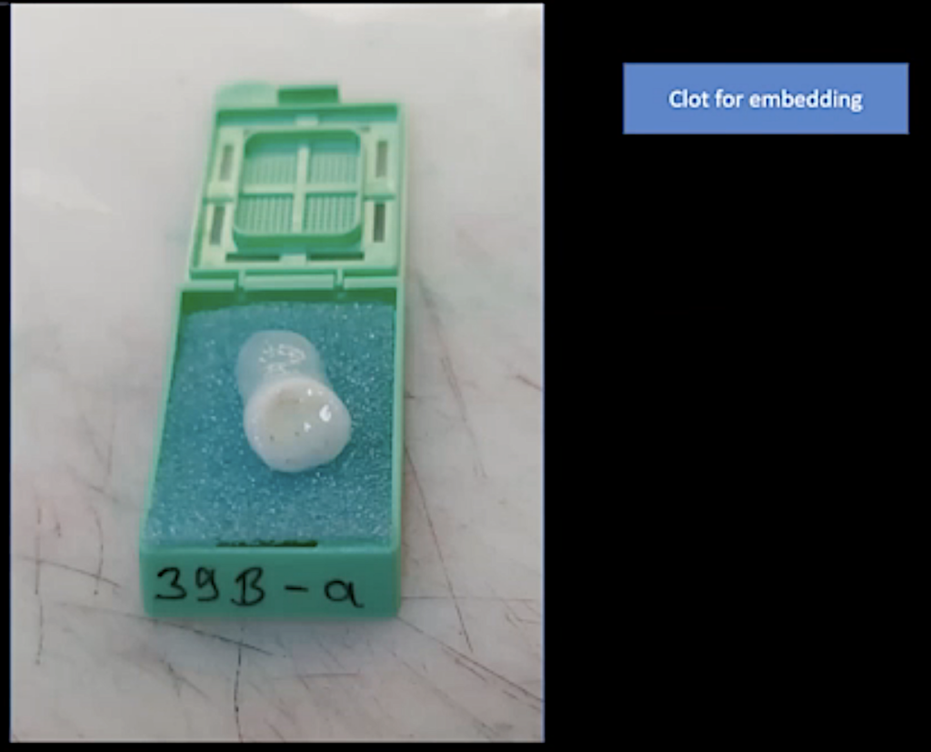
And we were asked, ‘Well, will you examine these clots formed by this lady after the cooling?’ and we did this.
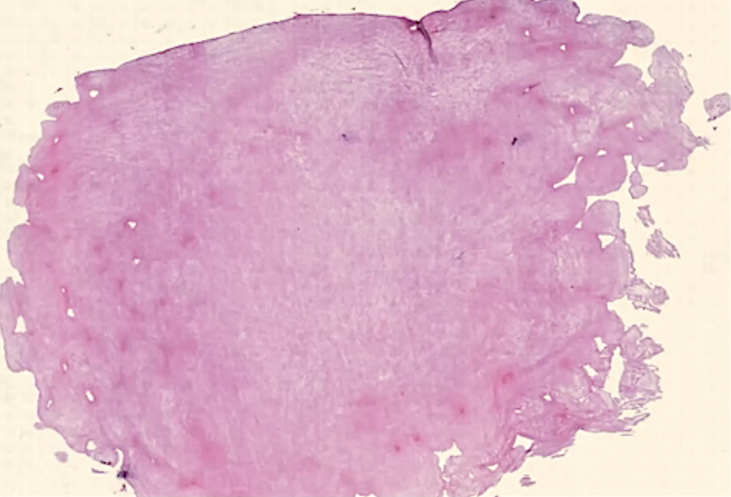
Here you can see this is the clot that we took, and you can see the histology, it’s a proteinaceous-fibrous material which seems to be gross outgrowths on the periphery.
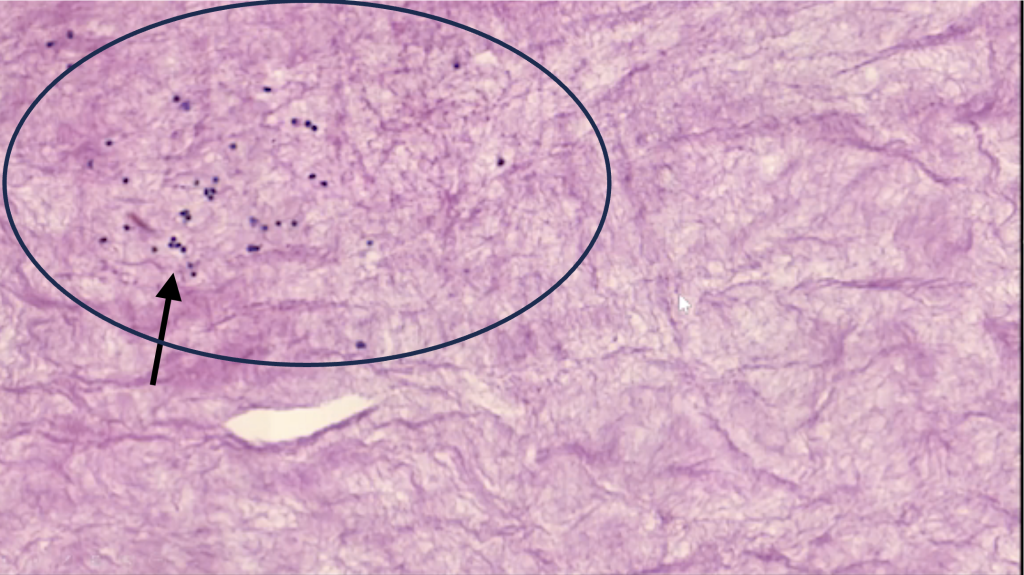
And there are some lymphocytes in there. It’s not mature fibrin but fibrinogen, and we did some examination on it.
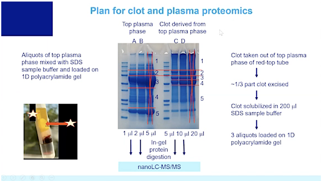
And you can see the top plasma phase and the clot derived from the plasma phase.
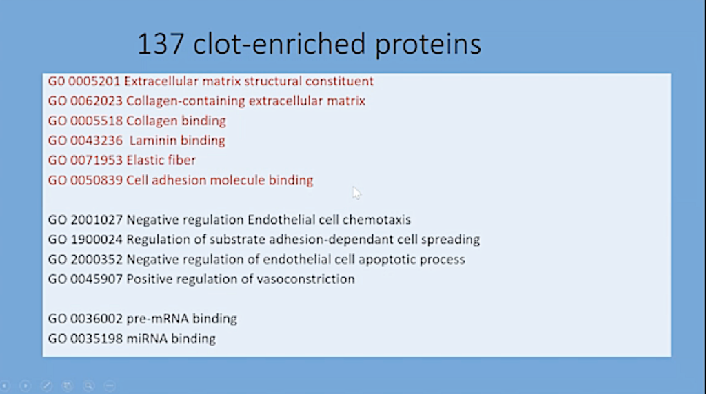
And there were 137 clot enriched proteins that were not found in the serum. These are all elements from the:
- Vessel walls,
- Extracellular matrix collagen,
- Collagen-containing extracellular matrix,
- Collagen binding,
- Laminin binding,
- Elastic fiber, and
- Cell adhesion binding molecule.
So, the conclusions that were drawn from this finding is that the endothelial damage persisted in this unlucky lady.
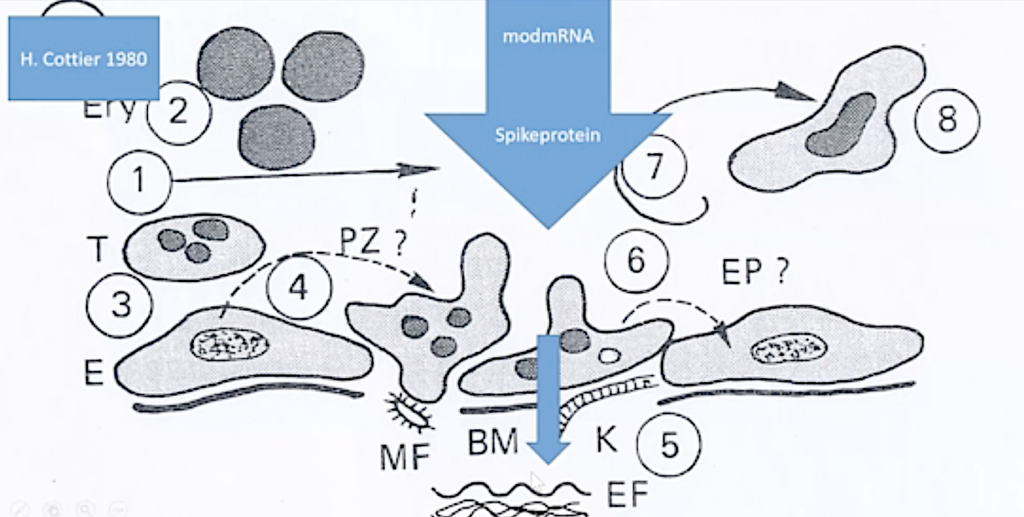
Here you can see a scheme. This is the endothelium, and, if the endothelium is destroyed by the spike protein, the spike protein goes to the deeper layer. And these constituents reach into the blood.
So, I think this is demonstration that the endothelial damage is very important and may persist for a very long time.
Specific Organ and Tissue Lesions (See SR 7.)
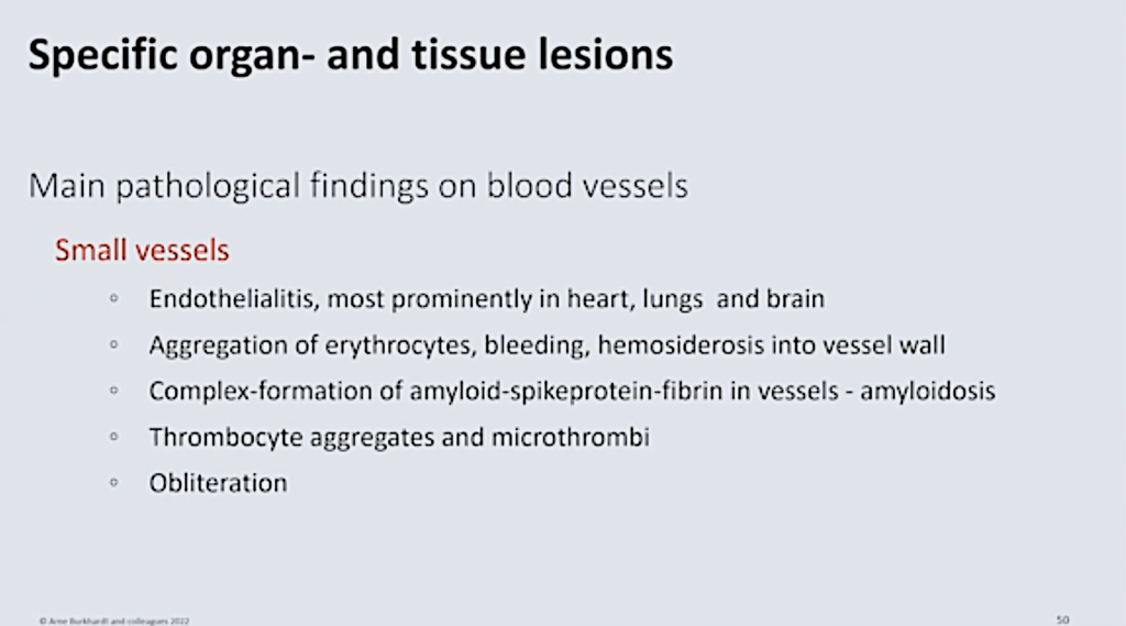
We come to the specific organ and tissue lesions, and we start with the main pathological findings in the blood vessels, the small vessels.
I already showed you the
- Endotheliitis most prominent in the heart, lungs and brain,
- Aggregation of erythrocytes,
- Hemorrhage and bleeding,
- Hemosiderosis into the vessel wall,
- Complex formation of amyloid-spike protein-fibrin in the vessel walls,
- Amyloid-like deposits,
- Thrombocyte aggregates, and, finally,
- Obliteration of small vessels.
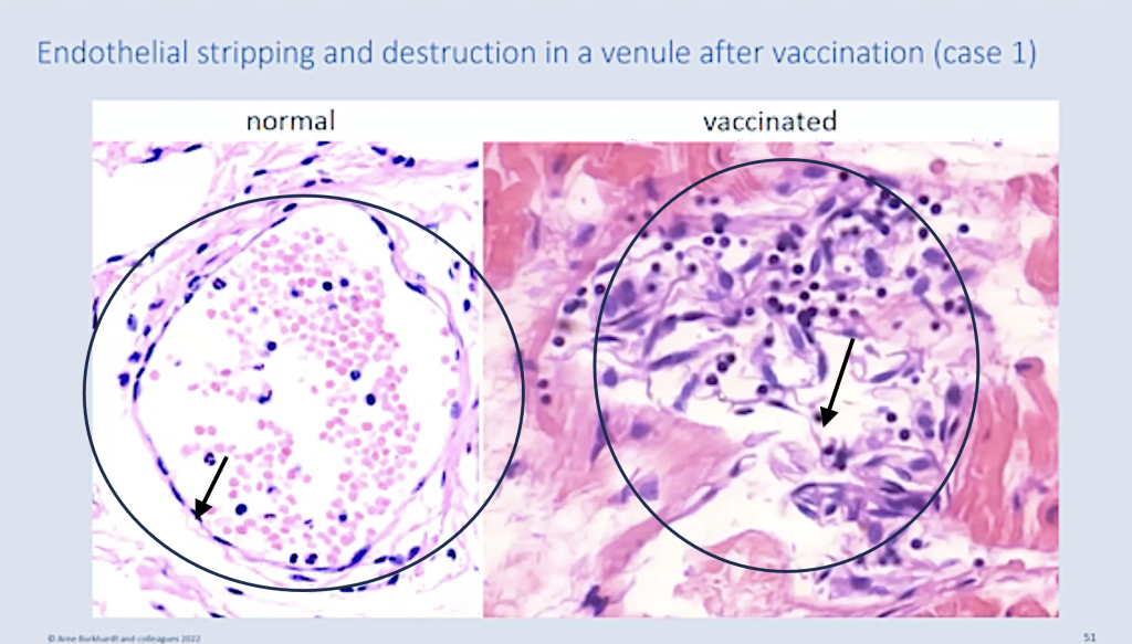
And here you can see the normal small vessels with the very thin endothelium, left. And here you can see a destroyed small vessel in the heart muscle, right. And you can see these spindle-like cells, these you can find here, these are the destroyed endothelium (arrow); and you can see the lymphocytic infiltration (oval), which clearly marks this as an intravital process.
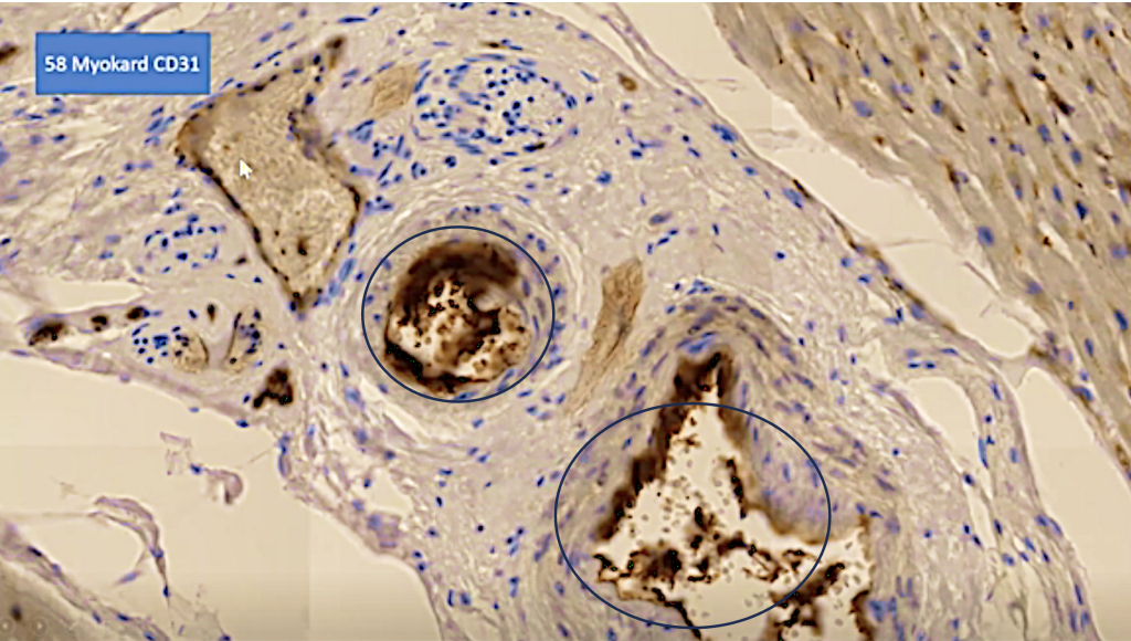
And we can show that these swollen and detached elements are endothelium by the CD 31 marker. (Cell adhesion molecule required for leukocyte transendothelial migration under most inflammatory conditions https://www.pathologyoutlines.com/topic/cdmarkerscd31.html.)
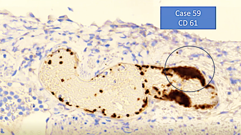
And then we can see that in these areas where the endothelium is destroyed there is attachment of platelets shown by the CD 61. (This glycoprotein complex (GPIIb-IIIa) binds plasma proteins, such as fibrinogen, fibronectin, von Willebrand factor, and vitronectin, and plays a critical role in platelet aggregation.)
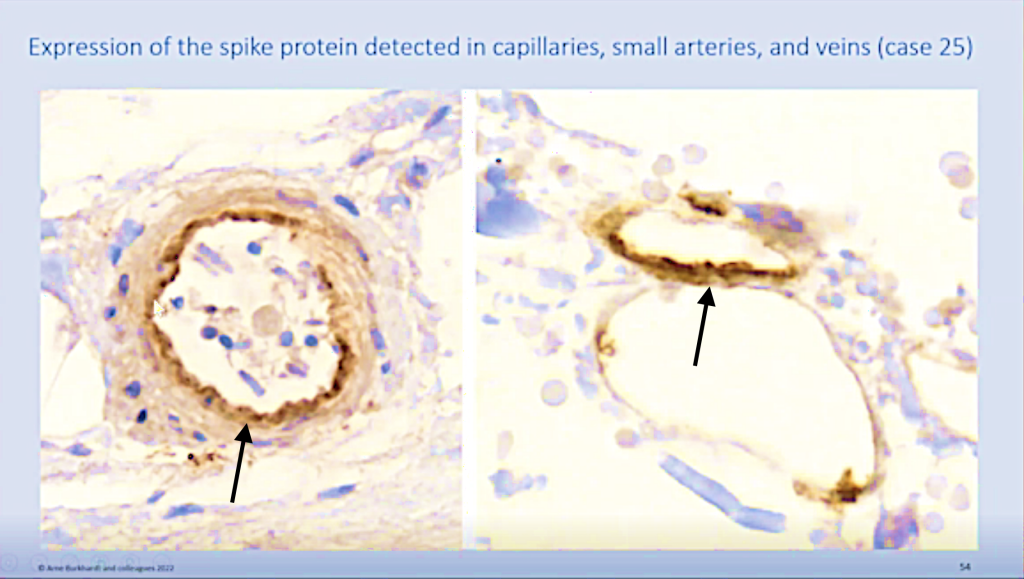
Of course, again, we can show the spike protein in these capillaries and in the small arterial vessels, not only in the endothelium, but also in the near inner most of the vessel walls.
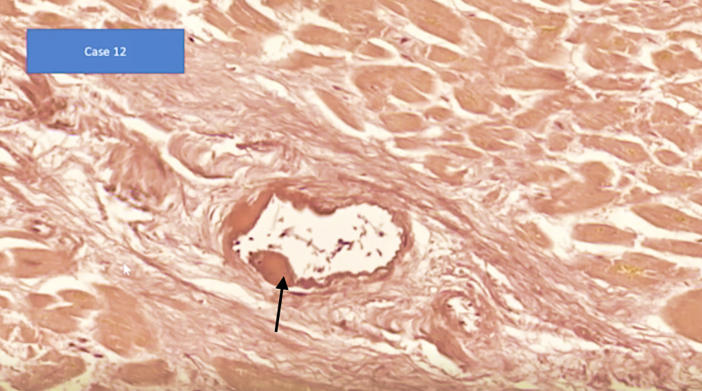
And, this can lead to these deposits here (black arrow).
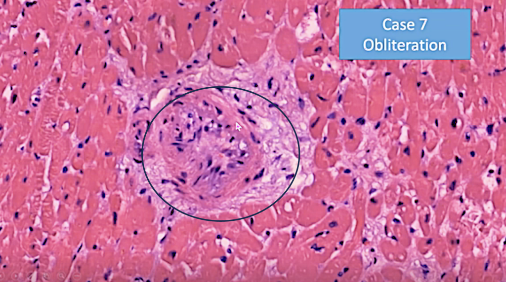
And finally, into obliteration here in the cardiac vessel…
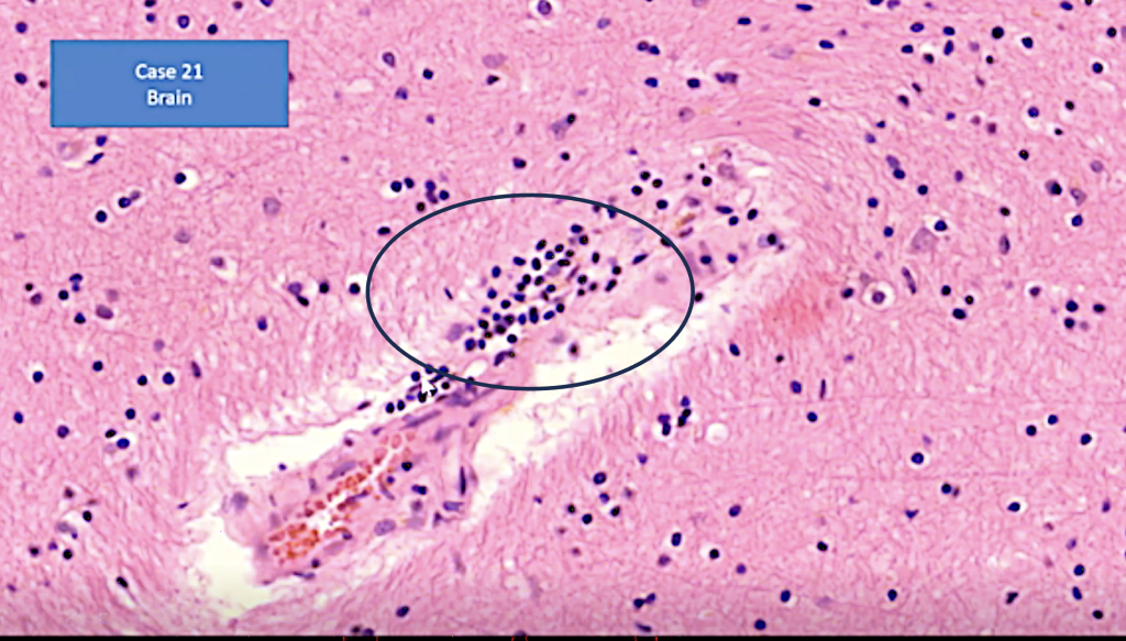
…also, inflammation of the small vessels in the brain. I will go to the brain later in detail, but this is in this context and…
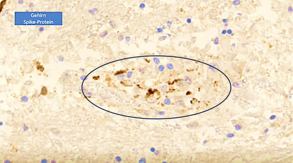
…you can see that in these vessel walls we can demonstrate the spike protein.
Large Vessels
Now, most alarming, is the finding in the large vessels.
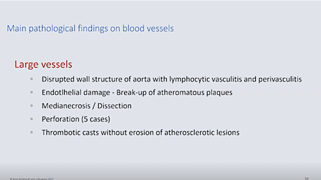
We see disrupted wall structure of the aorta with lymphocytic vasculation, vasculitis, and perivasculitis.
Again, the endothelial damage seems to be the leading adverse effect.
And, as I have also showed you, that this may lead to the breakup of atheromatous plaques.
Then in the deep media, we find necrosis and dissection.
Finally, perforation in five cases and thrombotic casts without erosion.
Aorta Aneurysm and Rupture (See SR 8.)
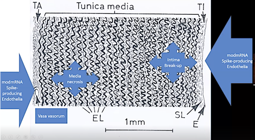
Now, again, this is a scheme of the order. And you can see there’s a two-front war, so to say, by the spike protein. It’s attacking the endothelium of the intima, and this may lead to break up. And they are attacking the endothelium of the vasa vasorum. And this apparently leads to media necrosis.
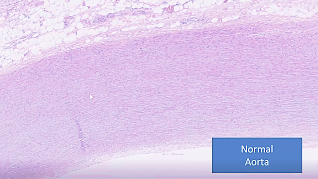
The normal aorta is just a very regular layers of elastic fibers and muscle cells. But here we observe media necrosis, and when do we find media necrosis or mesaortitis? It is with aortic rupture, like we have found in many, in so many of our cases. Now, it may be idiopathic, but this is usually, it’s a genetic defect, but it does not go with inflammatory.
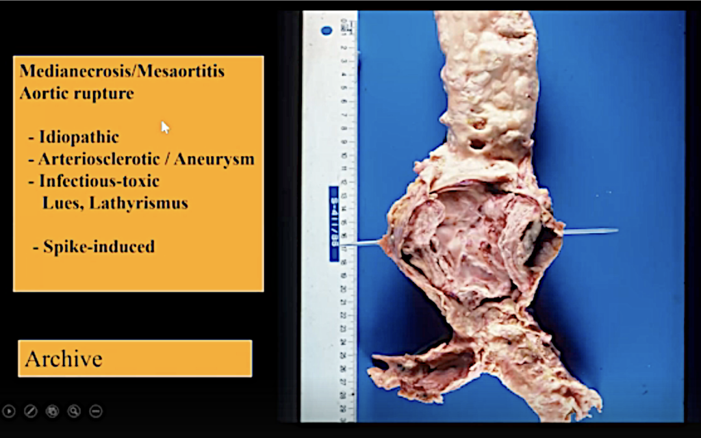
Then we have the atherosclerotic dissection, as we see here.
And finally, we have the toxic which we observe in Lues (late stage syphilis) and Lathyrism which is a rare poisoning from fava beans.
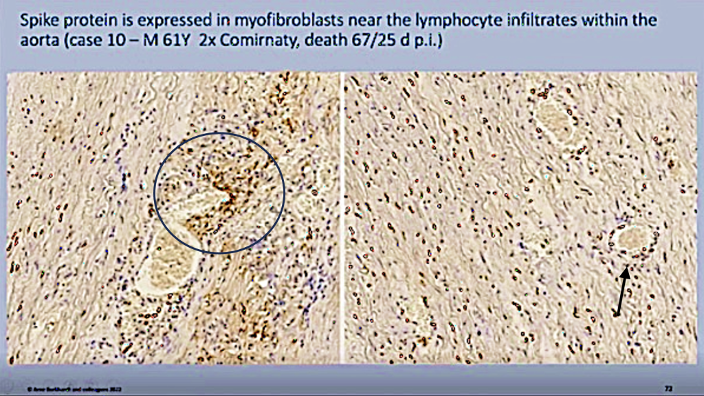
In our cases, apparently it is spike induced in these cases.
We could demonstrate the spike protein in the myofibroblasts of this damaged aorta (circle), and you can see mostly around vasa vasorum (black arrow) we found, we found this inflammatory infiltrate positive and here the myofibroblasts that are positive.
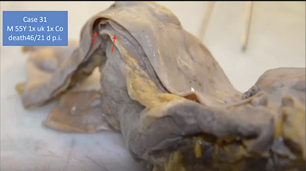
And this is just to show you how this looks in the organ specimen, and then you can see this split (red arrows), which is going all here; and you can see it’s, here the blood would be flowing, and you can see that this aorta is split in half, and this man died of the ruptured aorta.
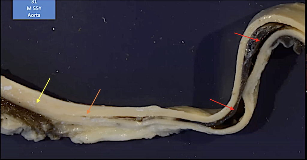
And you can see here (yellow arrow), here it’s still, the aorta is still intact in some areas, but here it starts to split (orange arrow). And then you can see the black, this is the blood (red arrows) that has formed here.
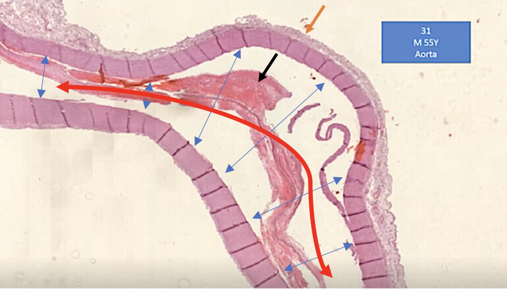
And in the tissue section you can see this also. The aortic wall is split (blue arrows) in two. And the blood flow would have, would have been in the path of the split wall (red arrow) of the aorta rather than where it should flow. This is the surrounding (orange arrow vasa vasorum). Here you can see the bleeding in this lesion (black arrow).
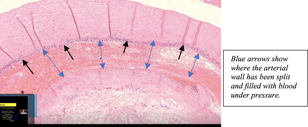
In this case, they thought it might be an idiopathic (possible genetic versus unknown causes) media necrosis; but, in contra distinction to the genetic media necrosis, we found a dense lymphocytic and macrophagic inflammatory infiltrate (black arrows), which apparently is induced by the spike protein toxicity.
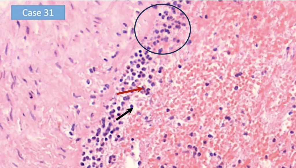
Here you can see a large magnification, there are lymphocytes (circle), some mast cells (red arrow) and some macrophages (black arrow). And here the bleeding has taken place.
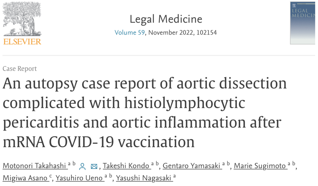
We are not the only ones who have seen this. This is in a report of a single case from Japan, just to mark this.
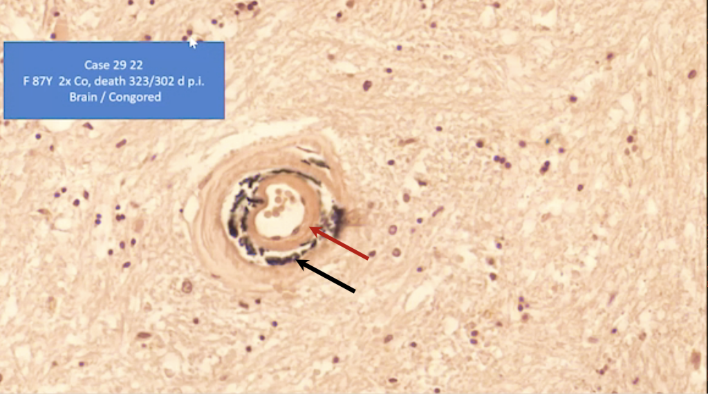
We can see the destruction of some part of the vessel walls, especially in many cases in the brain.
First of all, here you can see that there’s some deposition of amyloid-like (red arrow) substances at the innermost of the vessel.
And then you can see here where the elastic lamellae are situated (black arrow). They are destroyed, and they are strangely black.
We didn’t know at first what this was, but actually this is elastic lamellae incrustation, destroyed hemosiderin deposition caused by previous bleeding into the vessel wall.
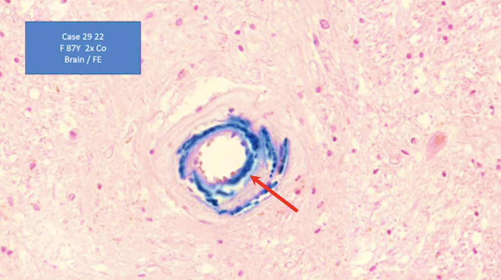
And here you can see it’s close to the artery that I showed you before, and you can see that these elastic lamellas are incrustated by hemosiderin (red arrow) as a residue of hemorrhage.
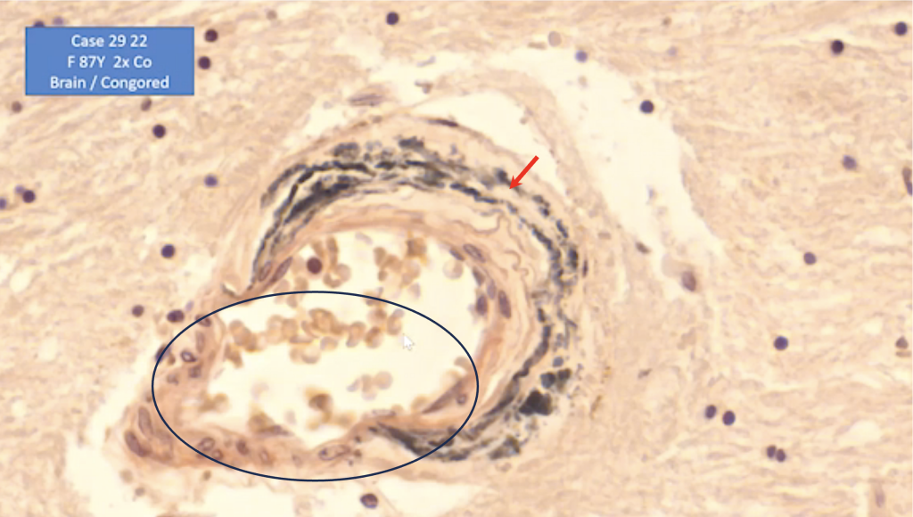
And here you can see again, these are the iron-positive, partly destroyed elastic lamella (red arrow). Here they are completely destroyed, and there is a micro-aneurysm which at any time might lead to bleeding, to fatal bleeding. (See SR 5.)
And we find this phenomenon not only in the arteries of the brain, but mostly in the brain.

But here you can see a larger artery in the thyroid gland which has these deposits of iron associated with the elastic lamella. (yellow arrows)
Sudden Adult Death Syndrome
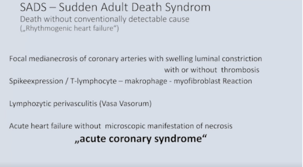
Now we consider that these lesions of the of the vessels may be one of the causes of the sudden death syndrome, a death without conventional detectable causes, which has not been known before this vaccination campaign. It usually is referred to as arrhythmogenic heart failure.
Now what we found might be the cause,
a) Focal media necrosis of the coronary artery and
i. swelling with luminal constriction,
ii. with thrombosis, or
iii. without thrombosis.
b) Spike expression, T lymphocyte macrophage, and myofibroblast reaction,
c) Lymphocytic perivasculitis. And this could be the cause of
d) Acute coronary syndromes without manifestation of an infection.
So, we come to the other main pathologic findings.
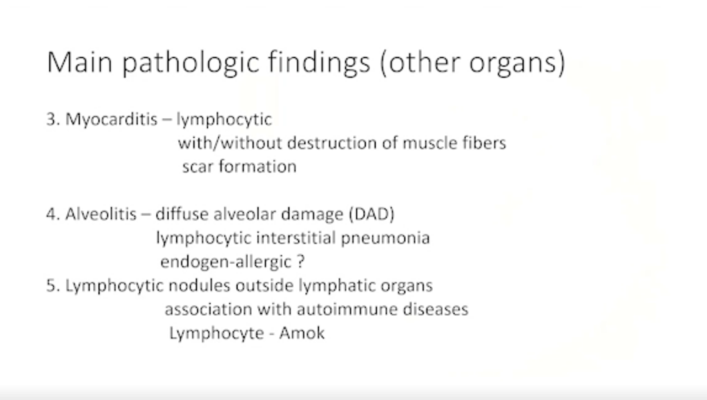
Myocarditis (See SR 9.)
We had the small and the large vessels, and we come to the myocarditis, which definitely is one of the most common findings. It’s lymphocytic. It is without destruction of most muscle fibers and, in some, and it leads to scar formation.
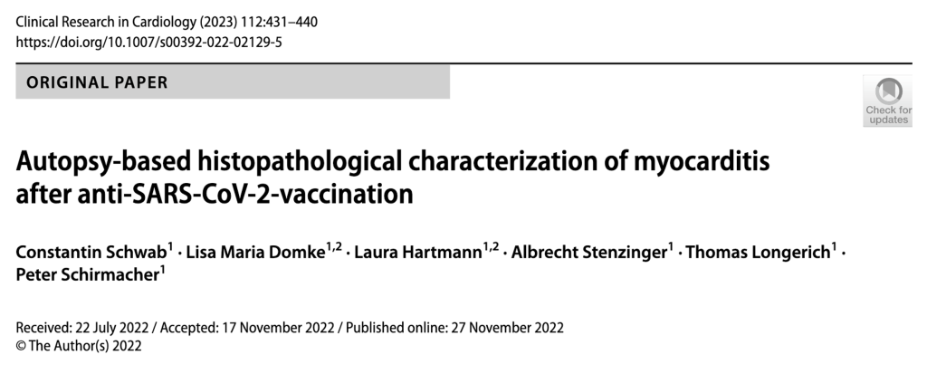
And this, as you may know now, is recognized by the international literature to be connected to this spike from the Corona vaccination. (Schwab, et al.)
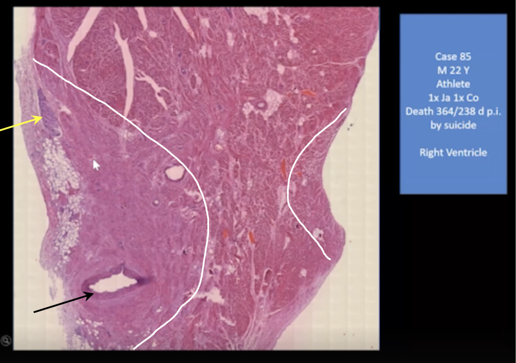
And here you can see a 22-year-old man, and he was an athlete; and he died by suicide because severe myocarditis was diagnosed.
And this is what we found at the autopsy.
- This is the left (right) heart chamber, and you can see he doesn’t have any pronounced arteriosclerosis (black arrow),
- Inflammatory infiltrates beneath the epicardium (yellow arrow).
- He has this large scar (white arrow and areas defined by white lines).
- Residual inflammatory infiltrates (yellow arrow).
This might be in the first injection was 364 days before he died. So, this could be formed after the first injection. The right side was in the healing process.
But in the left ventricle, it was still very active. And you can see these accumulations of lymphocytes (black circle).
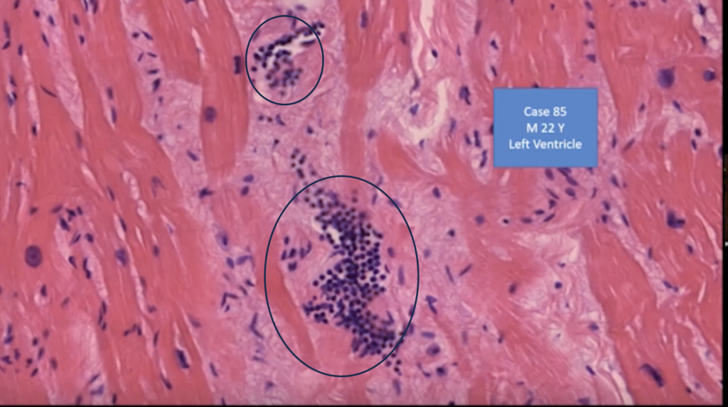
So probably in the consecutive injections, this inflammation of the myocardium was triggered again, boosted.
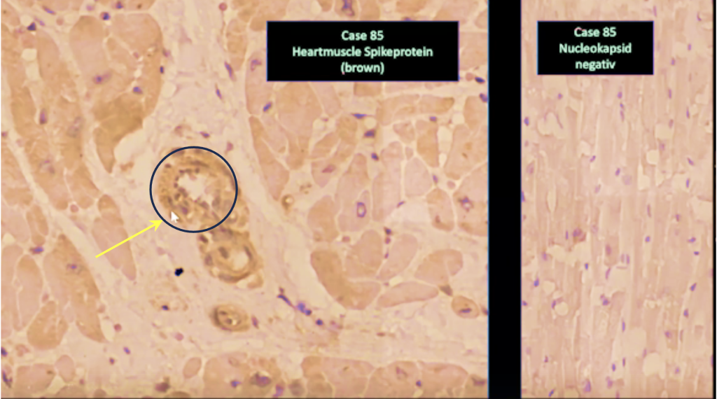
And in this case, you, we could show that the muscle cells expressed in variable amount the spike protein, but very pronounced (it was expressed) in the vessel walls of the small vessels. The vessel wall is swollen and has a disturbance of the structure (yellow arrow). We did the nucleocapsid stain in this case, and it was, as you can see, negative. (right image)
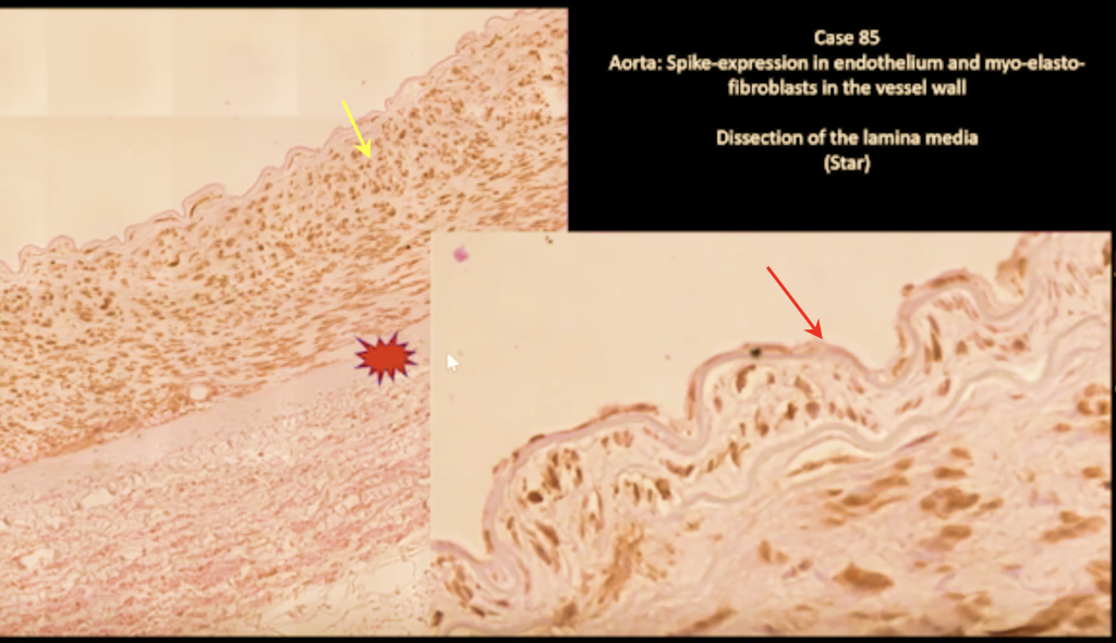
You can see here, this is the aorta, and you can see the endothelium here is intact (yellow arrow). You can see this clear demonstration of positive reaction for spike protein (red arrow). And you can see the dissection of the aorta (spiked red oval).
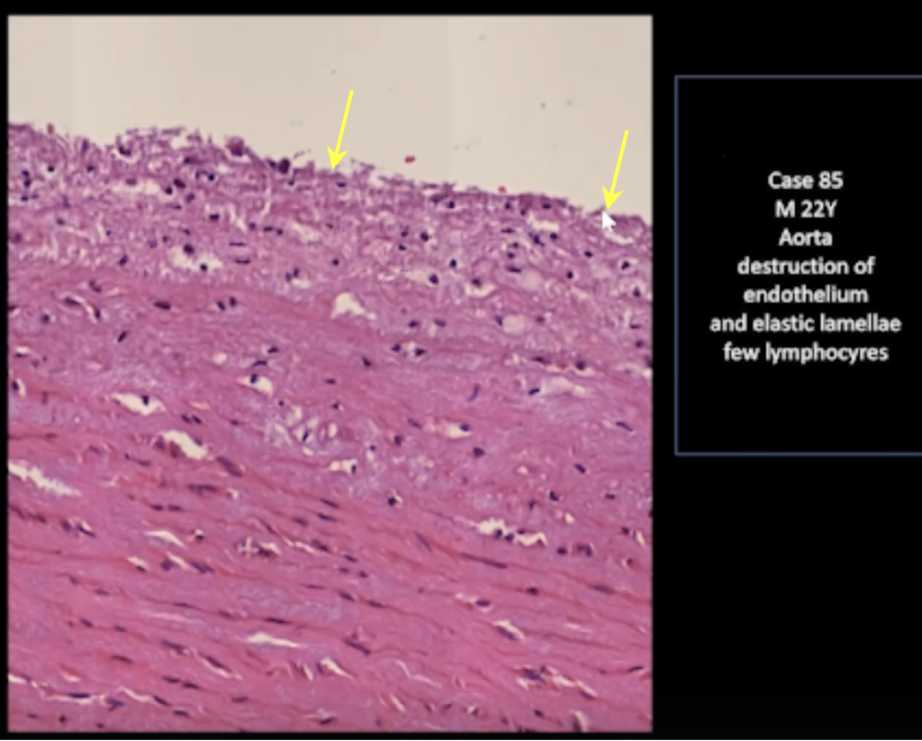
You can see another spot where the endothelium is destroyed (yellow arrows).
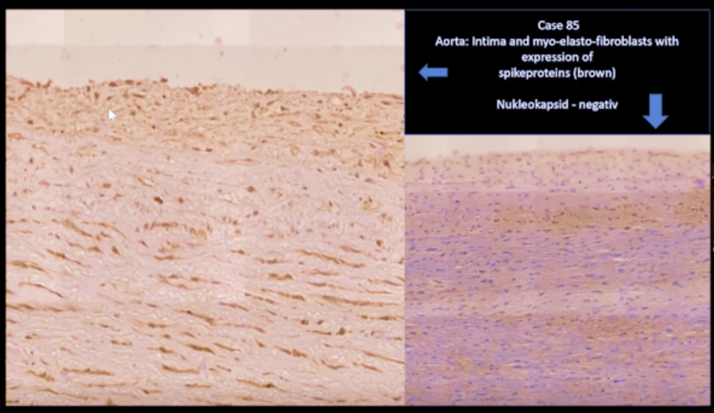
And here you can see the demonstration of spike protein and the negative control for nucleocapsid.
And, unfortunately, many doctors and medical persons state that the myocarditis usually is mild, and it is not of much concern.
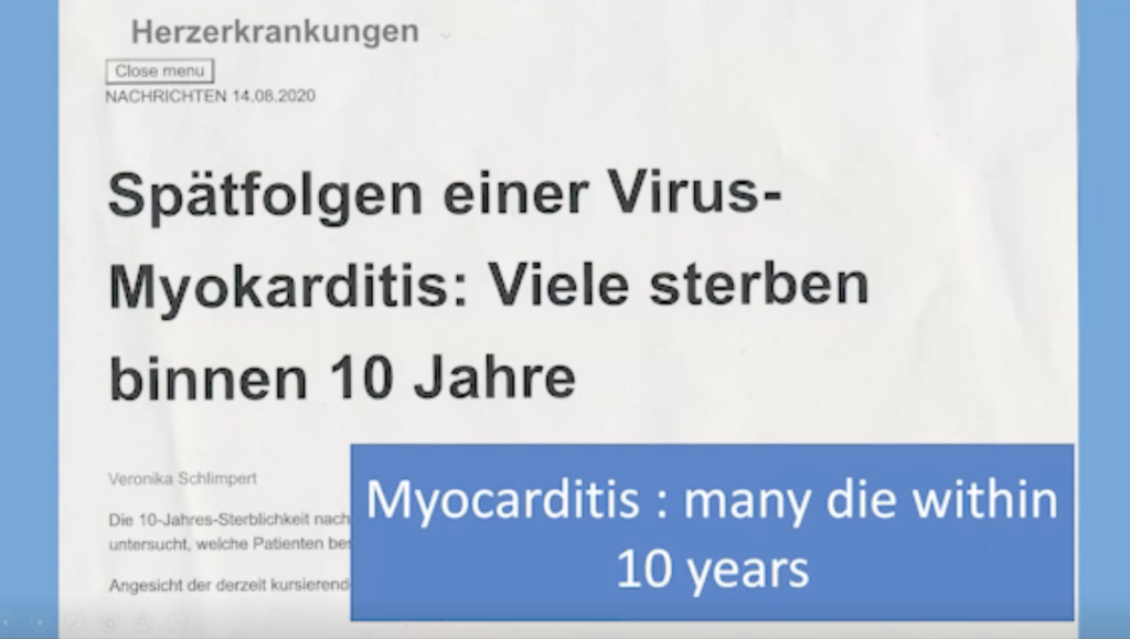
But this study here showed that many of the myocarditis patients die within 10 years, so it’s not something that should be taken easy.
Because, as an analysis by German cardiologists makes clear, the prognosis for viral myocarditis is generally quite unfavorable: almost 40% of the affected patients died within the next ten years, most of them from a cardiac cause, one in ten suffered sudden cardiac death. [Greulich, Simon, et al. “Predictors of Mortality in Patients With Biopsy‐Proven Viral Myocarditis: 10‐Year Outcome Data.” Journal of the American Heart Association, ahajournals.org, 13 Aug. 2020, www.ahajournals.org/doi/10.1161/JAHA.119.015351.]
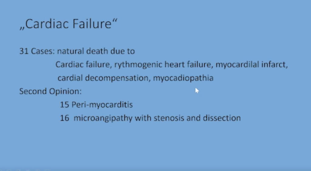
In our cases that were diagnosed as a natural cause, we had 31 cases where the primary pathologist or coroner said it was cardiac failure, they called it cardiac failure, arrhythmogenic heart failure, myocardial infarct, cardial decompensation, myocardiopathia.
But, of our second opinions, in 15 of these 31 cases there was a clear cut peri-myocarditis.
And in 60 cases we had this, what I showed you, microangiopathy with stenosis and dissection. So, this is one of the main causes of death after the vaccination.
Lung and Alveolitis
Now we come to the next organ, the lung.
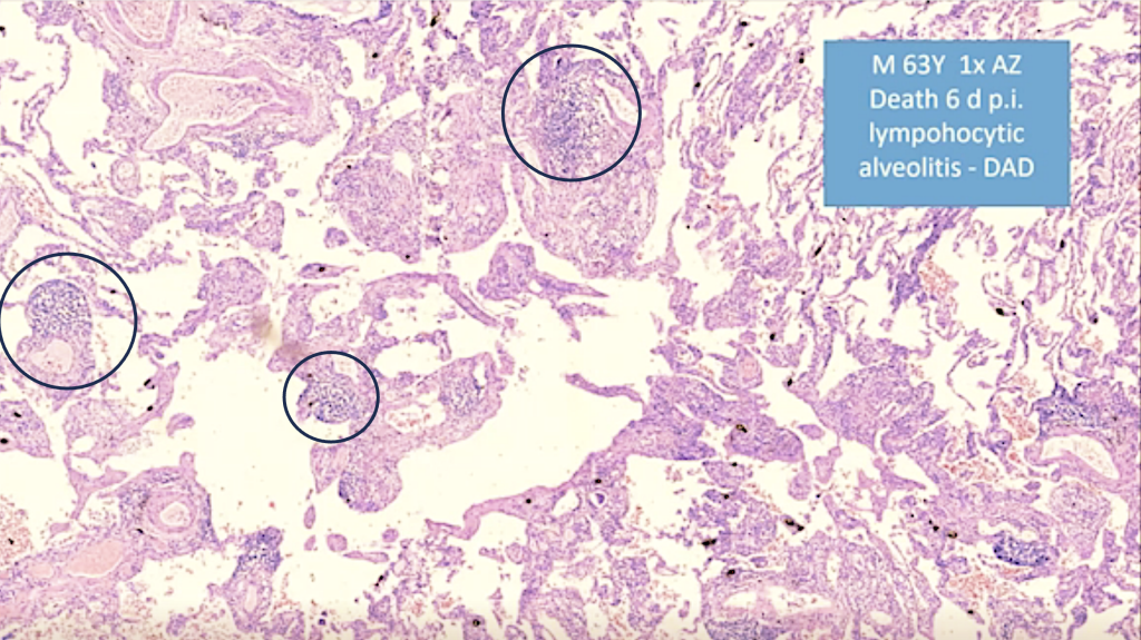
In the lung we have a lymphocytic alveolitis and focal infiltrations of lymphocytes, mainly perivascular and in some areas what we call diffuse alveolar damage.
Lymphatic Organs
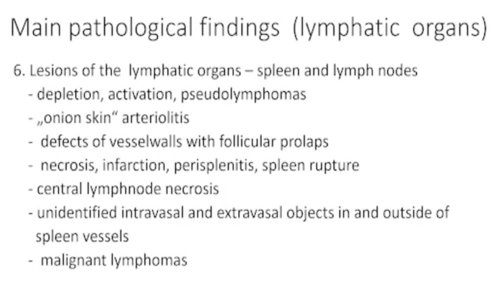
Now we come to the lymphatic organs, lesions of the lymphatic organs, spleen, and lymph nodes. We have,
- Depletion, activation, and pseudolymphomas,
- ‘Onion skin’ arteriolitis (typical of autoimmune diseases),
- Defects of the vessel walls with follicular prolapse,
- Necrosis, infarction, spleen rupture
- Central lymph node necrosis,
- Unidentified intravasal and extravasal objects.
- Malignant lymphomas.
One of the main organs where the vaccination and the spike protein is active is the spleen.
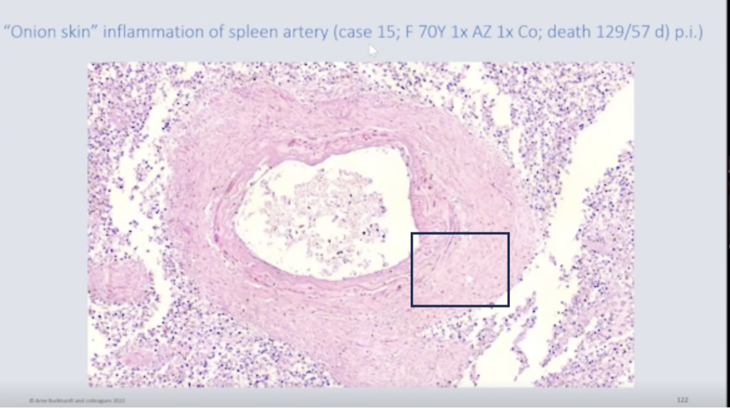
You can see this is what is called the onion skin-like changes of the vessel walls in the spleen, which, as I said, is, mainly seen in autoimmune diseases like lupus erythematosus.
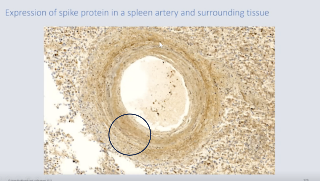
These disturbed small vessels also demonstrate the spike protein (small white arrow).
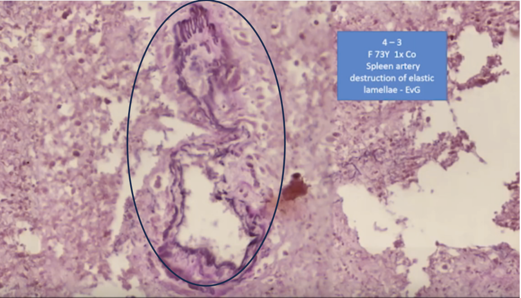
And here you can see some completely destroyed vessels. You can see the elastic lamella are completely destroyed.
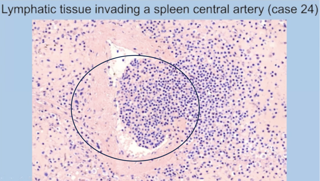
In some cases, the pressure of the proliferating lymphocytic follicles makes a disruption into these vessel walls.
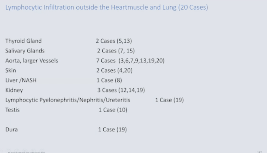
Autoimmunity and lymphocytic infiltration outside the heart muscle and lung

And here you can see where we find these lymphocytic infiltrates, thyroid, gland, salivary, aorta, skin, liver, kidney, testis, dura. And in these cases, we definitely observed autoimmune disease, Hashimoto’s, thyroiditis in five cases, Sjogren’s syndrome in four cases and atypical lichen planus with vasculitis in four cases.
Now, of course, this could be preexisting autoimmune diseases, but, apparently, they were not known before the deaths and before vaccination; and they might have been aggravated.
Now we come to the brain.
Brain
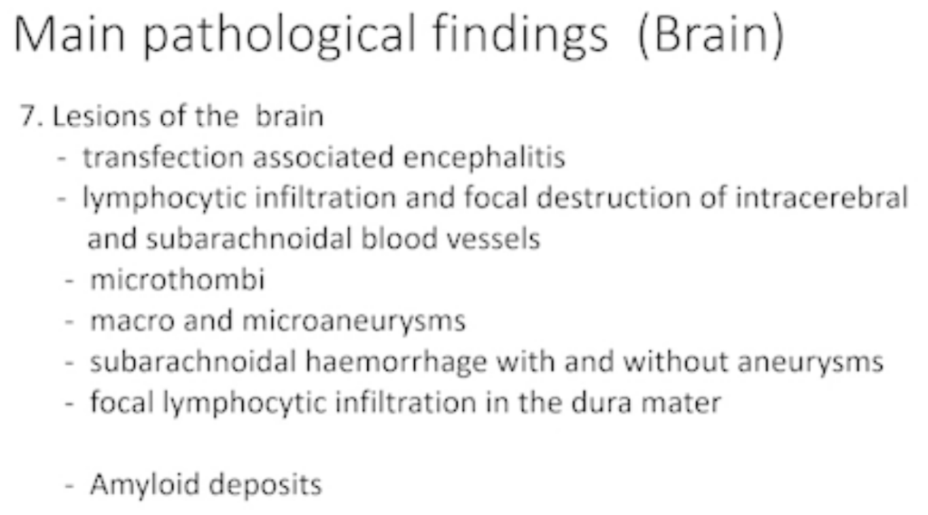
In the order of our main pathological findings, we have in a few cases,
- Transfection associated encephalitis,
- Lymphocytic infiltration and focal destruction of intracerebral and subarachnoid blood vessels,
- Microthrombi,
- Macro and microaneurysms,
- Subarachnoid hemorrhage with and without aneurysms,
- Focal lymphocytic infiltration in the dura matter,
- Amyloid deposits.
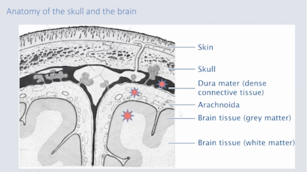
And this is the scheme where we have the lesions. We have the lesions, as I showed you, in the dura mater, where we have lymphocytic infiltrations. We have the arachnoidia. And we have the brain tissue. And, finally, I will show you this case report of this man to illustrate the findings in the brain. (See SR 5.)
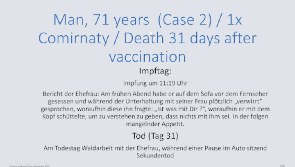
This is a man, 71-year-old. He was vaccinated one time with Comirnaty and died 31 days after this vaccination. But it’s very interesting that on the day he was… Oh, excuse me. This is in German. I have to translate it.
On the day of the vaccination, he fainted shortly and was disturbed and could not speak, but he recovered. So probably had, he had some lesion in the brain; but then he had recovered, and he worked in the in the in the forest and to cut down trees.
And when he made a break and sat in the car, suddenly he died unexpectedly.
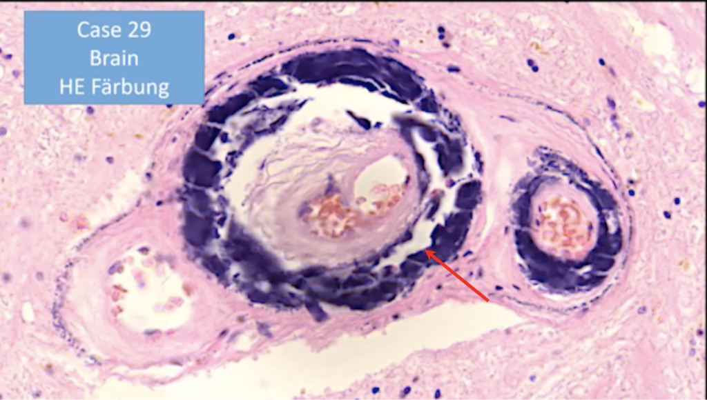
And this is what we found in the brain. This is a small artery, as you can see. There’s a dissection in the media (red arrow), and these elastic lamellae are destroyed, and they are strangely black stained by H&E.
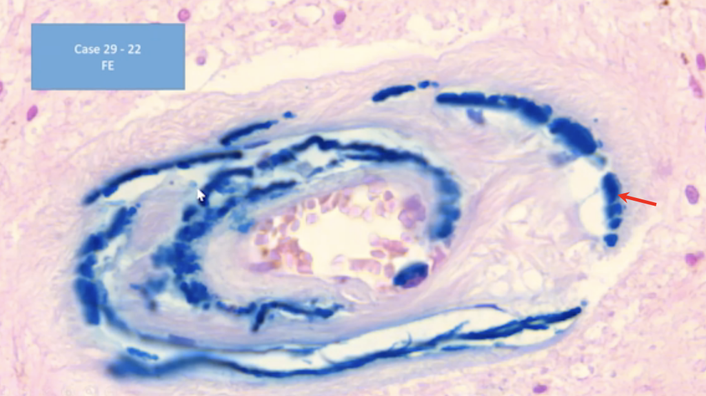
And as I have shown you before, this is all hemosiderin (red arrow), so he had probably on the day that he was vaccinated this bleeding inside the small vessels in the brain but recovered; and, days later, he had the fatal bleeding in the brain and died.
Pituitary Gland (See SR 10.)
So, finally, I just want to make one remark to the pituitary gland, which is situated near the brain, as you know, and has a very important function for the endocrine system, the same as the heart for the for the blood circulation. Now I found some very disturbing fact that of these 75 cases, only in eight cases the pituitary gland was examined by histology.
And also, when you look into the literature, in practically all of the reports that I read, the pituitary glands had not been examined, neither in the cases of COVID-19 infection, nor in the cases after vaccination. Now, the only public publication on this the subject was by Fitzek et al. And they found that, in some cases, there was necrosis of the pituitary.
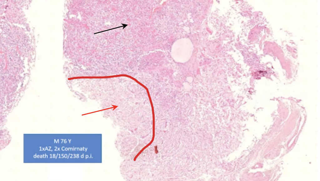
In one case, we could also find partial necrosis (red demarcation and arrow). This is an anterior lobe of the pituitary gland. Here it’s intact (black arrow), and here you can see the necrosis. So, this is definitely one thing that we should go further into.
So, I tried to show you our results, our slides and our findings. Of course, this is an ongoing study, and we don’t have the final answers to all of what we found or what I showed you. And I will be glad to discuss all of this with you.

Dr. Burkhardt passed away the day before his 80th birthday. We are fortunate that Dr. Burkhardt was able to add immeasurably to our understanding of the many manifestations of harms from spike-producing drugs.


June 22, 2023
“It’s Human Responsibility” – In Memory of Prof. Dr. Arne Burkhardt
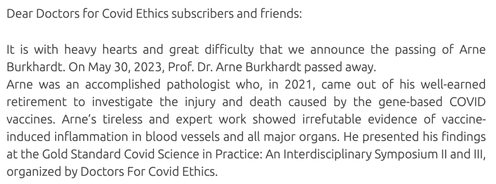
This tribute to Dr. Burkhardt contains three videos lasting 29:09, 41:14, and 1:12 minutes.
https://doctors4covidethics.org/its-human-responsibility-in-memory-of-prof-dr-arne-burkhardt/
Supplemental Resources
Links to these instructional videos are provided for background purposes. None are specific to COVID-related drugs or CoVax (COVID Vaccine-related) Diseases.
Part I: Pathological Processes
SR 1 Central Dogma in the Life Sciences
SR 2 Immune System Overview
SR 3 Inflammation after Injury
SR 4 Inflammatory Process (-itis)
SR 5 Cytotoxicity
SR 6 Amyloidosis
Part II: Specific Organs
SR 7 Vascular Disease and Vasculitis (Endotheliitis)
SR 8 Aneurysm
SR 9 Cardiac Scarring
SR 10 Pituitary and Hormones


